
- Title
-
The nucleoside diphosphate kinase NME3 associates with nephronophthisis proteins, and is required for ciliary function during renal development
- Authors
- Hoff, S., Epting, D., Falk, N., Schroda, S., Braun, D.A., Halbritter, J., Hildebrandt, F., Kramer-Zucker, A., Bergmann, C., Walz, G., Lienkamp, S.S.
- Source
- Full text @ J. Biol. Chem.
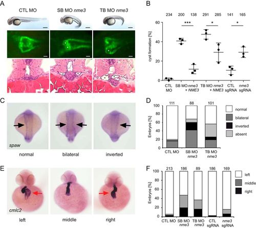
Knockdown of nme3 in zebrafish results in cyst formation and left-right asymmetry defects. A, zebrafish embryos injected with control (CTL) MO, nme3 SB MO, or nme3 TB MO at 48 hpf. In contrast to the control embryos, nme3 morphants showed a curly-tail phenotype. Scale bars, 100 μm. Nme3-depleted zebrafish embryos showed pronephric cyst formation, visualized using the transgenic Tg(wt1b:EGFP) line (white asterisks). Scale bars, 50 μm. Histological sections of nme3 morphants stained by hematoxylin and eosin confirmed cyst formation (black asterisks). Scale bars, 10 μm. B, quantification of the percentage of cyst formation caused by knockdown and CRISPR-mediated mosaic indel formation of nme3 at 48 hpf (*, p < 0.05; ***, p < 0.001; t test; error bars represent S.D.). C, representative pictures of 18-somite-stage zebrafish embryos after WISH for spaw with normal, bilateral, or inverted spaw expression (black arrows). D, quantification of the percentage of altered spaw expression in nme3 zebrafish morphants. E, examples of zebrafish embryos with normal (left) and reversed (middle and right) heart looping, visualized by WISH for the heart-looping marker cmlc2 at 48 hpf. The red arrows point to the atria. F, quantification of the percentage of heart laterality defects caused by nme3 depletion in zebrafish embryos at 48 hpf. EXPRESSION / LABELING:
PHENOTYPE:
|
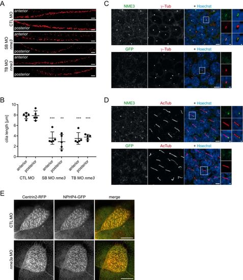
Nme3 is important for ciliogenesis in zebrafish and localizes to the ciliary basal body of mIMCD3 cells. A, confocal images of the anterior and posterior segments of the ciliated pronephric tubules of 24-hpf control (CTL) and nme3-deficient zebrafish embryos. Cilia were visualized by acetylated tubulin staining. Scale bars, 10 μm. B, quantification of the ciliary length in pronephric tubules. In contrast to the controls, ciliary length was affected by knockdown of nme3 (**, p = 0.0063; ***, p < 0.001; t test, error bars represent S.D.). C, confocal images of 6-day-starved mIMCD3 cells stained with antibodies against γ-tubulin (γ-Tub) (centrosomal marker; red), NME3 (green), and Hoechst (DNA; blue). NME3 colocalizes with γ-tubulin at the centrioles of centrosomes. D, costaining of cilia by acetylated tubulin (AcTub) (ciliary axoneme; red) identifies NME3 at the basal body of primary cilia. Immunofluorescence staining with an anti-GFP antibody serves as a control and results in the absence of centrosome and basal body labeling (bottom row). Scale bars, 5 μm. Magnifications and three-dimensional reconstructions are shown in white boxes. E, NPHP4–GFP and Centrin–RFP were coexpressed in epidermal multiciliated cells of stage 30 Xenopus embryos and detected by confocal microscopy. NPHP4 colocalization with the basal body marker Centrin was not altered by injection of nme3a morpholino in at least 25 cells observed in three experiments. Scale bars, 1 (C and D) and 10 μm (E). PHENOTYPE:
|

ZFIN is incorporating published figure images and captions as part of an ongoing project. Figures from some publications have not yet been curated, or are not available for display because of copyright restrictions. EXPRESSION / LABELING:
|