The evolution of cranial dermal bones and the developmental mechanisms. (A–C) The skull roof of Eusthenopteron(A), Acanthostega(B), Ichthyostega(C). (D) The developmental mechanisms of the frontal bone in mice. The molecules that initiate dermal differentiation from the ectoderm to the mesenchyme have not been identified yet, though the WNT signaling pathway is suggested to be involved (Goodnough et al., 2014). ano, anocleithrum; esl, lateral extrascapular; esm, medial extrascapular; f, frontal; ip, interparietal; n, nasal; o, opercular; oc, occipital; p, parietal; pf, postfrontal; po, postorbital; pop, preopercular; pot, post-temporal; pp, postparietal; prf, prefrontal; st, supratemporal; sq, squamosal; sclei, supracleithrum; t, tabular. Illustrations in (A–D) are redrawn with permissions from Clack (2012), Jarvik (1980), and White et al. (2016).

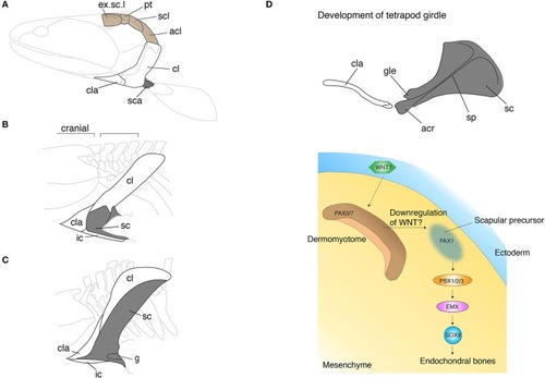
The skeletal shift from dermal to endochondral bones in the pectoral shoulder girdles. (A–C) The pectoral girdles of Eusthenopteron(A), Ichthyostega(B), Eryops(C). Brown shaded bones (extrascapular, post-temporal, supracleithrum, and anocleithrum) in (A) have been lost during the fish-to-tetrapod transition. Shaded bones depict endochondral bones. Note that the endochondral bones have enlarged with the concomitant decrease of the dermal bones. In (B,C), the spaces between the skull and pectoral girdle demonstrate the origin of the neck. (D) The developmental mechanisms of the scapula in mouse and chicken embryos. The epithelial-mesenchymal transition produces prospective scapular cells from the dermomyotome. The understanding of developmental programs underlying the dermal bone development is still poor (see text). acl, anocleithrum; acr, acromion; cl, cleithrum; cla, clavicle; g, glenoid fossa; ic, interclavicle; sc, scapula; sca, scapulocoracoid; scl, supracleithrum; sp, spine; Illustrations in (A–C) are redrawn with permissions form Andrews and Westoll (1970), Jarvik (1980), and Gregory (1951). The illustration of the mouse scapula is adapted with permission from Kuijper et al. (2005).

The fin-to-limb transition and their developmental basis. (A–C) The pectoral fins and limbs of Eusthenopteron(A), Tiktaalik(B), and Acanthostega(C). Note that Eusthenopteron and Tiktaalik possess distal fin rays with the endochondral skeletons. (D) The developmental mechanisms of fin rays and digits. In zebrafish, hox13 genes are expressed in a distal domain of the endochondral disk. The cells that experienced the late phase hox activity migrate out from the distal endochondral disk into the fin fold, that differentiate into the lepidotrichia. In mice, Hox13 genes are expressed in the autopod, which develop digits at a later stage. HOXA13 is suggested to bind the regulatory region of Bmp2 and 4 by ChIP experiments (Knosp et al., 2004). While many of the downstream genes of HOX13 have been explored in mice, genes regulated by HOX13 in fish have not been identified. Note that the ossification of lepidotrichia and digits takes place at a later stage than the expression of Hox13 genes. (E) Hypotheses for the fin-to-limb transition. The fin rays (cells shaded by light orange) have degenerated and the endochondral domain (cells shaded by light gray) expanded during the appendage evolution (top). This hypothesis supports that digits and wrists are novel domains that have been acquired as fish have evolved to tetrapods. Another hypothesis claims that the fish fin has an antecedent of digits and wrists (bottom). The cell histories between fish fin rays and tetrapod digits are comparable in terms of their hox expression pattern during the embryonic development (cells shaded by orange). In this hypothesis, the cell differentiation program of fin rays might have changed to become endochondral bones, resulting in the acquisition of digits and the wrist. dr, distal radials; hu, humerus; int, intermedium; ir, intermediate radials, le, lepidotrichia; po, postaxial process, pr, preaxial radials; pro, proximal radials; ra, radius; ul,ulna, uln, ulnae; Illustrations of (A–C) are redrawn with permissions from Andrews and Westoll (1970), Shubin et al. (2006), and Coates (1996).

Acknowledgments
This image is the copyrighted work of the attributed author or publisher, and ZFIN has permission only to display this image to its users. Additional permissions should be obtained from the applicable author or publisher of the image. Full text @ Front Cell Dev Biol