
- Title
-
Foxi1 promotes late-stage pharyngeal pouch morphogenesis through ectodermal Wnt4a activation
- Authors
- Jin, S., Jiyun, O., Stellabotte, F., Choe, C.P.
- Source
- Full text @ Dev. Biol.
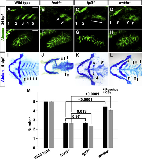
Requirement for Foxi1 in the late-stage of pouch development. (A-H) Alcama immunohistochemistry (green) showed five pouches (1–5 in A) in wild-type embryos at 34 hpf. foxi1 mutants had posterior pouches shaped abnormally (arrows in B), whereas fgf3 mutants had two anterior pouches (1 and 2 in C) with a cell mass that failed to migrate out to form posterior pouches (line in C). wnt4a mutants displayed four abnormally shaped pouches (arrows in D). Scale bar: 40 µm (A-D). (E) Alcama immunohistochemistry (green) showed a normal bilayered pouch morphology in wild-type embryos. (F and H) In foxi1 and wnt4a mutants, an aberrant, similar multilayered pouch morphology was found. (G) In fgf3 mutants, posterior pouch-forming cells failed to migrate out. Scale bar: 20 µm (E-H). (I-L) Ventral views of facial cartilages. In wild-type embryos, a bilateral set of five CBs (arrows in I) formed. Fewer CBs were observed in foxi1 and wnt4a mutants, whereas fusions of CBs (arrowheads in K) were observed with reduced anterior CBs (asterisks in K) in fgf3 mutants. (J, L) Normally shaped CBs were marked with arrows in foxi1 and wnt4a mutants. (M) Quantification of pouch and CB defects in wild-type embryos and mutants. Data represent mean ± s.e.m. P values are shown. |
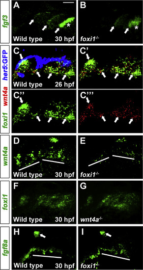
Foxi1 requirement in the ectodermal Wnt4a expression during pouch formation. (A, B) Fluorescent in situ hybridization showed fgf3 expression (green) in the posteriormost three pouches (arrows in A), with increased expression in the newly forming pouch (asterisk in A) in wild-type embryos. While fgf3 expression was observed in the posteriormost two pouches (arrows in B), the intensity in the newly forming pouch (asterisk in B) was weaker than that of wild-type embryos (compare to asterisk in A). (C) Double fluorescent in situ hybridization showed colocalization of wnt4a (red) with foxi1 (green, arrows in C) but not with her5-positive pouch endoderm labeled by GFP immunohistochemistry (blue). (C’) Green and red channels. (C”) Green channel only. (C′′′) Red channel only. (D, E) Fluorescent in situ hybridization for wnt4a (green). In the facial ectoderm, foxi1 mutants had significantly reduced wnt4a staining, compared with wild-type embryos (lines in D and E). (F, G) Fluorescent in situ hybridization for foxi1 (green). Similar foxi1 expression in the pharyngeal regions was observed in wnt4a mutants (G), compared with wild-type embryos (F). (H, I) Fluorescent in situ hybridization for fgf8a (green). (H) In wild-type embryos, fgf8a was expressed in mesoderm (line) adjacent to outgrowing pouches as well as in the otic vesicle (arrow). (I) In foxi1 mutants, fgf8a expression was observed in the mesoderm (line) as well as in the otic vesicle (arrow), even though fgf8a expressing mesoderm was disorganized compared to wild-type mesoderm. Scale bar: 40 µm. |
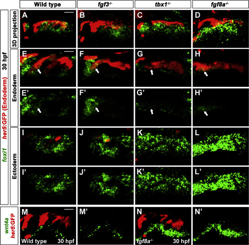
Requirements for Tbx1 and Fgf8a in repression of the ectodermal Foxi1 and Wnt4a. (A-L) Fluorescent in situ hybridization for foxi1 (green) and GFP immunohistochemistry to detect her5:GFP-positive endoderm (red) at 30 hpf. (A, B) In wild-type embryos and fgf3 mutants, foxi1 expression was observed in her5:GFP-positive endoderm (arrows in E and F) as well as in facial ectoderm (I and J). (C) In tbx1 mutants, foxi1 expression was significantly decreased in the her5:GFP-positive endoderm (arrow in G), whereas it was expanded ectopically in the facial ectoderm (K). (D) In fgf8a mutants, foxi1 expression was detected in the her5:GFP-positive endoderm (arrow in H) and foxi1 expression in the facial ectoderm was expanded ectopically and increased in intensity (also see L). (E-G) Higher magnification image of wild- type embryos and mutants focusing on the her5:GFP-positive endoderm. (I-L) Higher magnification image of wild-type embryos and mutants focusing on the facial ectoderm. (M, N) Fluorescent in situ hybridization for wnt4a (green) and GFP immunohistochemistry to visualize her5:GFP-positive pouches (red) at 30 hpf. wnt4a expression was observed in ectodermal patches in wild-type embryos (M), whereas it became stronger and was expanded ectopically in the facial ectoderm of fgf8a mutants (N). (E′-N′) Green channel only. Scale bars: 40 µm (A-D, M, and N), 20 µm (E-L). |
Reprinted from Developmental Biology, 441(1), Jin, S., Jiyun, O., Stellabotte, F., Choe, C.P., Foxi1 promotes late-stage pharyngeal pouch morphogenesis through ectodermal Wnt4a activation, 12-18, Copyright (2018) with permission from Elsevier. Full text @ Dev. Biol.