
- Title
-
Genetic and developmental origins of a unique foraging adaptation in a Lake Malawi cichlid genus
- Authors
- Conith, M.R., Hu, Y., Conith, A.J., Maginnis, M.A., Webb, J.F., Albertson, R.C.
- Source
- Full text @ Proc. Natl. Acad. Sci. USA

µCT data demonstrate reduced ligament volume in adam12 zebrafish mutants. (A) Reconstructed 3D model in a wild-type zebrafish illustrating position of the cerato-mandibular ligament (cml) relative to mandible (den) and ceratohyal (ch). Maxilla (mx), premaxilla (pmx), and interopercle (iop) are also shown for perspective. (B) Reconstructed 3D model of the same structures in an adam12 mutant zebrafish. Note the marked reduction in the size of the cml (blue) and the coronoid process of the mandible (arrowheads). |
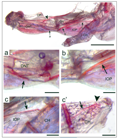
Direct red staining of an adult AB WT zebrafish jaw. Direct red stains both ligaments and tendons, making them easy to see under light microscopy. In this preparation, the certatohyal-mandibular ligament (CML) can be seen extending from the medial surface of the dentary bone (DNT) just below the coronoid process (CP) anteriorly (a), over the dorsal edge of the interopercle bone (IOP) (b & c), and onto the lateral surface of the ceratohyal bone (CH) posteriorly (c’). This preparation verifies the reconstruction of the CML from CT scans (Fig. 6 in the main text). In the main panel at top, the arrows indicate the path of the CML, the arrowhead shows the tendinous insertion of the second subdivision of the adductor mandibulae onto the dentary bone, the letters a-c correspond to the regions shown at higher magnification, and c’ shows the lateral face of the posterior region of the CH where the CML inserts. The abbreviation QU refers to the quadrate bone. The scale bar equals 1mm for the top panel. In panels a-c’ the scale bars all equal 200μm, the arrows point out the CML, and the arrowhead in c’ shows the insertion of the CML onto the CH. A note on the naming of the CML: Our use of this name is purely descriptive, as there is no mention of this ligament in the literature describing soft tissue anatomy in zebrafish. A CML has been described for damselfishes, and it serves an important role in feeding in this group, however whether these ligaments are homologous remains unknown at this time. |