FIGURE SUMMARY
Title

Evaluating the Capacity of Human Gut Microorganisms to Colonize the Zebrafish Larvae (Danio rerio).

Authors
Valenzuela, M.J., Caruffo, M., Herrera, Y., Medina, D.A., Coronado, M., Feijóo, C.G., Muñoz, S., Garrido, D., Troncoso, M., Figueroa, G., Toro, M., Reyes-Jara, A., Magne, F., Navarrete, P.
Source
Full text @ Front Microbiol

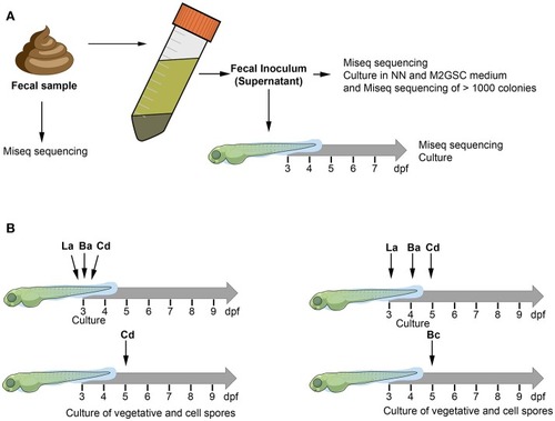
The figure illustrates the two experimental approaches used to humanize the zebrafish larvae. The composition of bacterial microbiota was analyzed through culture and MiSeq sequencing (A) Fecal sample was homogenized with pre-reduced PBS (0.1% cysteine) and allowed to stand for 5 min. The supernatant of the homogenized solution was used as inoculum to transplant 3 dpf zebrafish larvae. (B) Zebrafish larvae (3, 4, or 5 dpf) were inoculated by immersion with a suspension of each bacterium at a final concentration of 108 CFU/mL in sterile E3 medium. La: Lactobacillus acidophilus, Ba: Bifidobacterium adolescentis, Cd: Clostridioides difficile, Bc: Bacillus clausii.

Macroscopic and microscopic morphology of vegetative cells and endospores of (A)Bacillus clausii and (B)Clostridioides difficile. Upper show bacterial colonies in TSA or NN media. Middle show DTAF fluorescent bacteria. Lower show green malachite staining with black arrows indicating green endospores.

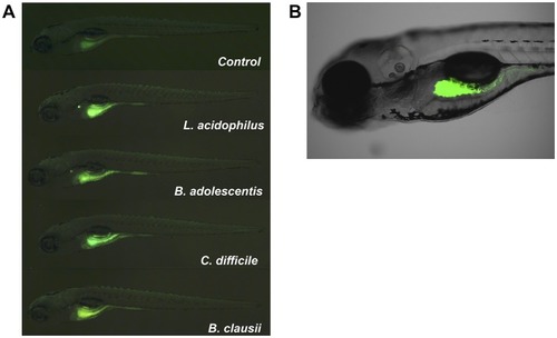
Localization of fluorescent bacteria (DTAF) in 5 dpf zebrafish larvae. Observation of larvae in an (A) SZX16 stereoscope (Olympus) with a Micro Publisher 5.0 RVT camera (QImaging) and (B) confocal laser scanning microscope Olympus FluoView FV1000 Spectral, Software version 2.1. The figure shows different larvae after 2 h of being inoculated with DTAF-labeled bacteria.

Acknowledgments
This image is the copyrighted work of the attributed author or publisher, and ZFIN has permission only to display this image to its users. Additional permissions should be obtained from the applicable author or publisher of the image. Full text @ Front Microbiol