
- Title
-
MicroRNA-194 Regulates the Development and Differentiation of Sensory Patches and Statoacoustic Ganglion of Inner Ear by Fgf4
- Authors
- Cao, H., Shi, J., Du, J., Chen, K., Dong, C., Jiang, D., Jiang, H.
- Source
- Full text @ Med. Sci. Monit.
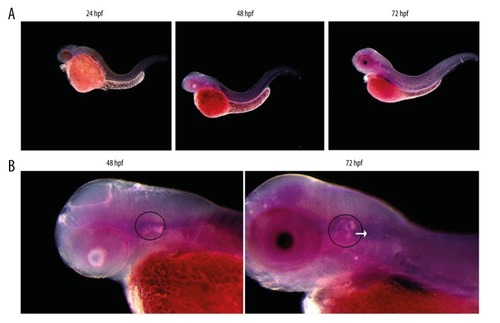
Expression of miR-194 in the embryos and inner ear of zebrafish. (A) Expression of miR-194 in the embryos of zebrafish from 24 to 72 hpf. (B) Expression of miR-194 in the inner ear of zebrafish. Inner ear was indicated by a black circle; the VCG was indicated by a white arrow. VCG – vestibulocochlear ganglion; hpf – hour post fertilization. EXPRESSION / LABELING:
|
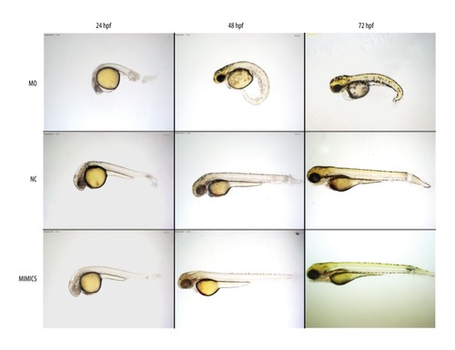
Altered expression of miR-194 affected the development of embryos from 24 to 72 hpf. In the MO group, the whole development of embryo slowed down, the edema of the pericardium, the small head, and the axis of the body were skewed. But there was no obvious change in the whole development and inner ear development pattern in the mimics group. MO – microinjection with morpholino oligonucleotide; NC – negative control; Mimics – microinjection with miR-194 mimics; hpf – hour post fertilization. PHENOTYPE:
|

Effect of altered expression of miR-194 on the development of inner ear and otolith. (A) Effect of altered expression of miR-194 on the development of inner ear from 24 to 72 hpf. A black circle was used to indicate inner ear. (B) Changing the expression of miR-194 affected the otolith. MO – microinjection with morpholino oligonucleotide; NC – negative control; Mimics – microinjection with miR-194 mimics; hpf – hour post fertilization. PHENOTYPE:
|
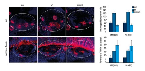
MiR-194 promoted the differentiation of the sensory patches in zebrafish inner ear. The percentage of Sox2 (upper portion) and acetylated-tubulin (lower portion) positive cells were measured by in situ hybridization at 48 hpf. The white circle area indicated zebrafish inner ear. Red staining indicated Sox2 expression (upper portion) or acetylated-tubulin expression (lower portion); blue indicated cell nucleus. MO – microinjection with morpholino oligonucleotide; NC – negative control; Mimics – microinjection with miR-194 mimics; hpf – hour post fertilization. PHENOTYPE:
|

MiR-194 promoted the development and differentiation and regulation of the normal spatial structure of SAG in zebrafish inner ear. The percentage of Islet1 (upper portion) and HuC (lower portion) were measured by in situ hybridization at 48 hpf. The white circle area indicated zebrafish inner ear. Red staining indicated Islet1 expression (upper portion) or HuC expression (lower portion); blue indicated cell nucleus. MO – microinjection with morpholino oligonucleotide; NC – negative control; Mimics – microinjection with miR-194 mimics; hpf – hour post fertilization; SAG – statoacoustic ganglion. PHENOTYPE:
|

MiR-194 regulated the development and differentiation of inner ear sensory patches by modulating the expression of Fgf4. The percentage of Sox2 (left panel) and acetylated-tubulin (right panel) were measured by in situ hybridization at 48 and 72 hpf. The white circle area indicated zebrafish inner ear. Red staining indicated Sox2 expression (left panel) or acetylated-tubulin expression (right panel); blue indicated cell nucleus. MO – microinjection with morpholino oligonucleotide; NC – negative control; MO+FGF4 – microinjection with morpholino oligonucleotide and Fgf4; hpf – hour post fertilization. PHENOTYPE:
|
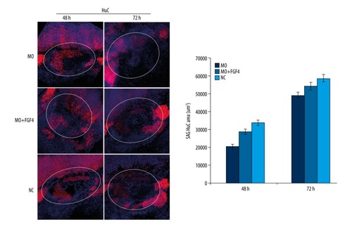
MiR-194 regulated the development and differentiation of SAG in inner ear by regulating the expression of Fgf4. The percentage of HuC were measured by in situ hybridization at 48 and 72 hpf. The white circle area indicated zebrafish inner ear. Red staining indicated HuC expression; blue indicated cell nucleus. MO – microinjection with morpholino oligonucleotide; NC – negative control; MO+FGF4 – microinjection with morpholino oligonucleotide and Fgf4; hpf – hour post fertilization. PHENOTYPE:
|