
- Title
-
Microvillar and ciliary defects in zebrafish lacking an actin-binding bioactive peptide amidating enzyme
- Authors
- Kumar, D., Thomason, R.T., Yankova, M., Gitlin, J.D., Mains, R.E., Eipper, B.A., King, S.M.
- Source
- Full text @ Sci. Rep.
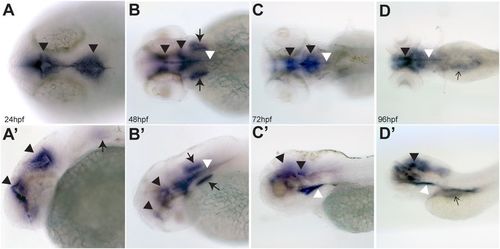
PAM expression in zebrafish embryos. Dorsal (A–D) and lateral (A’–D’) views of zebrafish embryos at 24, 48, 72 and 96 hpf visualized by in situ hybridization with an antisense probe for pam; staining with the sense control probe using the same processing conditions is shown in Supplemental Fig. S1. pam mRNA expression is detected in the ependyma lining the developing ventricles (black arrowheads), otic vesicles (thick arrows), floor plate (white arrowheads), and was also diffusely abdominal (thin black arrows in panels D and D’). No pam expression was evident in the eye or cardiac tissue. |

ZFIN is incorporating published figure images and captions as part of an ongoing project. Figures from some publications have not yet been curated, or are not available for display because of copyright restrictions. |

Phenotypic analysis of pam−/− zebrafish embryos. Dorsal and lateral views of wildtype (panels A,C,E,G,I,K and M) and pammbg5−/− (panels B,D,F,H,J,L and N) embryos; comparison of wildtype (A and C) and pammbg5−/− mutants (B and D) revealed no obvious differences at 24 and 48 hpf. By 72 hpf, pammbg5−/− embryos develop pericardial edema (asterisk in F); by 96 hpf, additional edema around the abdomen (thin arrows in H and J), irregular cyst-like protrusions in the abdominal region (inset in H), and smaller eyes (arrowheads in H and J) were observed. The general area of the pronephros is indicated by thick arrows (in H and L). Severe edema developed at 5 dpf in pammbg5−/− mutants (L and N). Additional edema in the brain was also visible (red arrowhead in L). Developmentally normal wildtype sibling controls at 5 dpf are shown for comparison (K and M). A key to the symbols used is provided. |
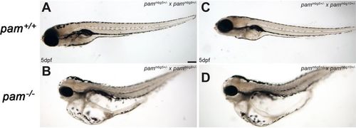
Heteroallelic pam−/− zebrafish display severe edema. Heterozygous zebrafish bearing different pammbg alleles were crossed and the phenotypes of the resulting pam−/− heteroallelic embryos and their wildtype siblings were examined at 5 dpf. Wildtype pam+/+ embryos developed normally (A,C). In contrast, heteroallelic pam−/− homozygotes (pammbg5/pammbg9 and pammbg5/pammbg10) derived from pammbg5+/− × pammbg9+/− and pammbg5+/− × pammbg10+/− crosses exhibited severe edema (B,D). PHENOTYPE:
|
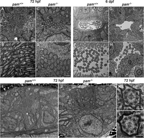
pam−/− zebrafish exhibit microvillar and ciliary assembly defects in the pronephros. Transmission electron micrographs of transverse sections through the pronephros of pam−/− embryos and their wildtype siblings at 72 hpf (A–D and I,J) and 6 dpf (E–H); the images shown were taken from approximately midway along the pronephros. At 72 hpf, the pronephros lumen of wildtype zebrafish is occluded with numerous cilia surrounded by a dense array of microvilli (A,B). Although densely packed cilia were evident in the pronephric lumen of pam−/− embryos at this stage, brush border microvilli were absent (C,D). At 6 dpf, the lumen of the wildtype zebrafish pronephros was more open; numerous cilia appeared in the lumen, which was surrounded by epithelial cells extending a dense array of microvilli (E,F). In contrast, the lumen of the pam−/− pronephros showed a severe reduction in the number of cilia along with the continued absence of a brush border (G,H). Occasional cytosolic axonemes (inset in H) and undocked centrioles/basal bodies (arrow in G and see Supplemental Fig. S6) were observed in the epithelial cells lining the pronephric duct of the pam−/− embryos. Lower magnification images (I,J) of the region surrounding the pronephros of the 72 hpf embryos shown in panels (A,B) illustrate the general cellular architecture. The cilia present in pam−/− embryos at 72 hpf were morphologically normal (K,L). Bars = 100 nm (K,L), 500 nm (A,C,F,H), 250 nm (B,D), 1 μm (G) and 2 μm (E,I,J). PHENOTYPE:
|

Dorsal (A) and lateral (A’) views of a wildtype 48 hpf zebrafish embryo subject to in situ hybridization using the pam sense probe, with the same photographic conditions as in Fig. 1. No signal was observed in the ventricles, otic vesicles or floor plate, confirming the specificity of the signals observed with the antisense probe shown in Fig. 1. (B) Dorsal view of a wildtype 24 hpf embryo subject to in situ hybridization with the pam sense probe; this image was acquired using DIC optics. No signal was observed in any tissue. |

Cilia are present in the olfactory bulbs, neuromasts and otic vesicles of pam-/- embryos Confocal immunofluorescence images of the olfactory bulb (A, B), neuromast mechano-sensory cells (C, D), and otic vesicles of control and pam-/- embryos probed with antibody against acetylated tubulin. Electron micrographs (G, H) revealed that at 6 dpf both kinocilia (KC) and actin-based stereocilia (SC) were evident emanating from the sensory hair cells in the otic vesicles of pam-/- embryos; these structures assemble early in development at ~16 hpf. The insets in (A, B) show that the olfactory motile cilia have normal 9+2 ultrastructure. Bars = 5 μm (A-F), 1 μm (G, H) and 100 nm (insets in A and B). PHENOTYPE:
|

Cyst-like structures and edema in 6 dpf pam-/- embryos Toluidine blue-stained transverse section through a 6 dpf pam-/- embryo. Massive edema was evident (*) as were cyst-like structures (arrows) which were routinely observed associated with the pronephric tubules. The boxed region in A is shown at higher magnification in B. Bars = 10 and 2.5 μm for panels A and B, respectively. PHENOTYPE:
|

Ciliary and microvillar defects in the pronephros of 6 dpf pam-/- embryos Upper panels - Electron micrographs of the anterior-posterior regions of the pronephros of 6 dpf pam+/+ (A-E) and pam-/- (F-J) embryos. The entire pronephros was examined using alternating 0.5 μm thick sections for ~ 50 μm followed by several ultrathin sections examined by EM. Although microvilli and cilia were present in the most anterior segment of the pronephros of both wildtype and Pam-null embryos, cilia were much reduced, and microvilli were essentially absent in other pronephric regions of the mutant; only the very posterior pronephros lacked microvilli in the wildtype embryo. Bars = 2 μm (A, E, F, J) and 1 μm (B, C, D, G, H, I). Lower panels – Bright-field micrographs of toluidine blue-stained transverse thick sections through wildtype (K, L) and Pam-null (M, N) 6 dpf embryos; the vastly different appearance of the mutant embryos is due to the massive edema. Higher magnification images of the pronephric region (L, N) revealed that the pronephros lumen (white arrows in L) in the pam+/+ embryo was almost occluded, whereas it was more open in the pam-/- animals. Bars = 10 (K, M) and 5 (L, N) μm. PHENOTYPE:
|

Undocked centrioles/basal bodies in 6 dpf pam-/- embryos Transverse sections through wildtype and pam-/- embryos (6 dpf). (A, B) Adjacent sections through a cell lining the pronephros of a 6 dpf wildtype embryo showing two basal bodies (BB) with striated rootlets (SR) docked at the plasma membrane with attached membrane-bound ciliary axonemes (cilium) extending into the pronephric lumen; numerous microvilli (MV) are evident. (C, D) Undocked centrioles/basal bodies (arrows) located within the cytoplasm of cells lining the pronephros of a 6 dpf pam-/- embryo and oriented approximately parallel to the pronephros long axis as are the cytosolic axonemes (see Fig. 5H); few cilia are present in the lumen and microvilli are absent. Bar = 500 nm. PHENOTYPE:
|