
- Title
-
The Beta-Adrenergic Agonist Salbutamol Modulates Neuromuscular Junction Formation in Zebrafish Models of Human Myasthenic Syndromes
- Authors
- McMacken, G., Cox, D., Roos, A., Müller, J., Whittaker, R., Lochmüller, H.
- Source
- Full text @ Hum. Mol. Genet.

ZFIN is incorporating published figure images and captions as part of an ongoing project. Figures from some publications have not yet been curated, or are not available for display because of copyright restrictions. |
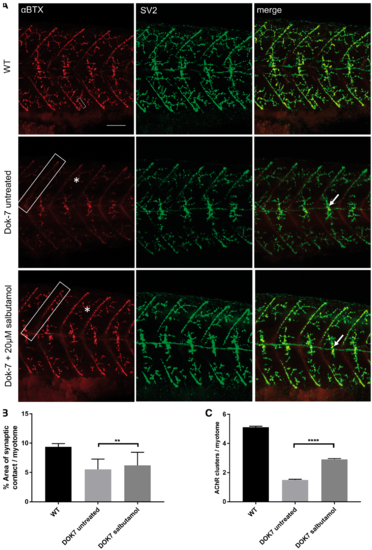
Salbutamol treatment of Dok-7 MO-injected zebrafish improves NMJ morphology. Lateral views of 48 hpf embryos with neuromuscular synapses labelled with antibodies against SV2 (green, presynaptic vesicles) and αBTX (red, postsynaptic AChRs). Scale bar = 50 µm. (A) In Dok7 MO-injected embryos, the focal innervation point along the horizontal myoseptum is reduced in size compared with wild type, but is increased following treatment with 20 µm salbutamol (arrows). In addition, the morphology of myoseptal (boxes) and myotomal (asterisks) AChR clusters is partially rescued with salbutamol treatment. A representative 20 μm2 AChR cluster is labelled with a bracket. (B) Following salbutamol treatment, the area of synaptic contact on the horizontal myoseptum is significantly increased (** indicates P < 0.01, Student’s t-test, n = 160 treated and 160 untreated myotomal segments examined). (C) Salbutamol treatment caused a significant increase in the number of AChR clusters >20 μm2 per myotome in Dok7 zebrafish embryos (**** indicates P < 0.0001, Student’s t-test, n = 100 treated and 100 untreated myotomal segments examined). PHENOTYPE:
|
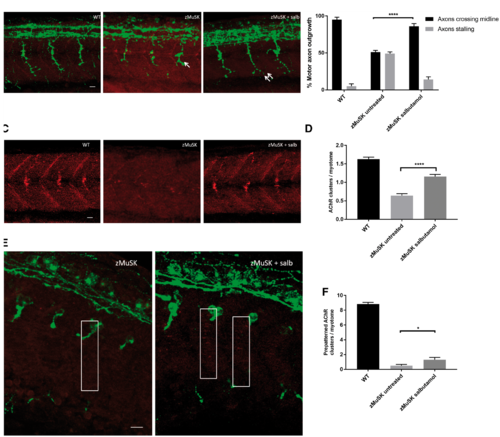
Salbutamol treatment of zMuSK MO-injected embryos improves motor axon pathfinding defects and AChR clustering. Lateral views of embryos with neuromuscular synapses labelled with antibodies against SV2 (green, presynaptic vesicles) and αBTX (red, postsynaptic AChRs). Scale bars = 10 µm. (A) zMuSK MO-injected embryos at 24 hpf demonstrate characteristic stalling of the outgrowing motor axon at the choice point on the horizontal midline (arrow). Following salbutamol treatment more axons cross the horizontal midline and extend to the periphery of the myotome (double arrow). (B) Quantification of the improvement of motor axon growth following salbutamol treatment. (**** indicates P < 0.0001, chi-square test, n = 240 treated and 240 untreated myotomal segments examined). (C) zMuSK embryos at 24 hpf display almost complete absence of AChR clustering. In salbutamol-treated zMuSK embryos, AChR clustering is restored. (D) Quantification of the improvement in AChR clustering following salbutamol treatment of zMuSK embryos. AChR clustering is significantly increased with the mean number of AChR clusters >20 μm2 per myotome 1.16 in salbutamol treated embryos compared with 0.64 in untreated embryos (**** indicates P < 0.0001, Student’s t-test, n = 100 treated and 100 untreated myotomal segments examined). (E) zMuSK MO-injected embryos at 17 hpf. Untreated embryos lack elongated, diffuse prepatterned clusters on the myotomal surface prior to the arrival of the motor growth cone (boxed areas), but following salbutamol treatment some zMuSK embryos display prepatterned clusters. (F) Quantification of the effect of salbutamol on prepatterned AChR clustering in zMuSK embryos. Following salbutamol treatment, the number of prepatterned AChR clusters >3 μm2 in zMuSK embryos increased, with a mean of 0.5 prepatterned clusters per myotome in untreated embryos, compared with 1.3 in salbutamol-treated embryos (* indicates P < 0.05, Student’s t-test, n = 30 untreated and 30 untreated myotomal segments examined). PHENOTYPE:
|
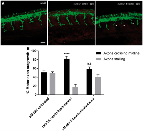
Pre-treatment with a selective β2 antagonist blocks the rescue of NMJ development by salbutamol. Lateral views of 24 hpf embryos with neuromuscular synapses labelled with antibodies against SV2 (green, presynaptic vesicles) and αBTX (red, postsynaptic AChRs). Scale bar = 50 µm. zMuSK embryos were treated with 5 µm of the selective β2 antagonist ICI118, 551 or control (water) added to E3 medium for 3 h prior to the addition of 20 µm salbutamol. (A) In zebrafish embryos pre-treated with ICI118, 551 no rescue of the axonal pathfinding defects was seen following 21 h of salbutamol treatment, with stalling and branching of motor growth cones (asterisks). (B) Quantification of the effect of pre-treatment of ICI118, 551 on motor axon growth. Following pre-treatment with 5 µm ICI118, 551, salbutamol treatment did not have an effect on axon pathfinding (n.s. P > 0.05, ****P < 0.0001, chi-square test, n = 240 treated and 240 untreated myotomal segments examined). PHENOTYPE:
|
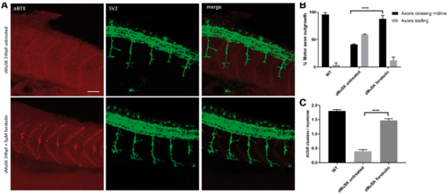
The effects of β2 receptor agonist activation on NMJ morphology are replicated by cAMP activation. Lateral views of 24 hpf embryos with neuromuscular synapses labelled with antibodies against SV2 (green, presynaptic vesicles) and αBTX (red, postsynaptic AChRs). Scale bar = 50 µm. (A) zMuSK embryos at 24 hpf demonstrate improvement in AChR clustering and axon pathfinding defects following forskolin treatment. (B) Quantification of motor axon guidance defects. About 88% (n = 28/240) of imaged somites of zMuSK embryos treated with forskolin displayed no axon guidance defects, compared with 41% (n = 142/240) of those in untreated zMuSK embryos. **** indicates P < 0.0001, chi-square test. (C) The number of AChR clusters larger than 20 μm2 on myotomal muscle fibres increased with forskolin treatment, with mean number of clusters 0.39 per myotome in untreated embryos, and 1.48 in treated zMuSK embryos. **** indicates P < 0.0001, Student’s t-test, n = 100 treated and 100 untreated myotomes examined. PHENOTYPE:
|