
- Title
-
Studying Diabetes Through the Eyes of a Fish: Microdissection, Visualization, and Analysis of the Adult tg(fli:EGFP) Zebrafish Retinal Vasculature
- Authors
- Wiggenhauser, L.M., Kohl, K., Dietrich, N., Hammes, H.P., Kroll, J.
- Source
- Full text @ J. Vis. Exp.
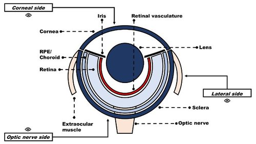
Schematic depiction of the adult zebrafish eye. |

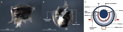
Removal of the adult zebrafish eye. Corneal side view with the eye still in the orbital cavity and intact optic nerve (A). Corneal side view with detached eye (B). Schematic depiction of the zebrafish eye at this step (C). |
Adult zebrafish eye with extraocular muscles in focus. Lateral view with attachment of an extraocular muscle to the left outer rim of the ocular globe in focus (A). Optic nerve side view with extraocular muscles symmetrically flanking the optic nerve (B). Schematic depiction of the zebrafish eye at this step (C). |

Adult zebrafish eye after removal of all extraocular muscles. Lateral view with the cleared outer rim of the ocular globe visible (A). Optic nerve side view with optic nerve in the middle. The light-reflecting sclera is not covering the whole area around the nerve (red dashed line) (B). Schematic depiction of the zebrafish eye at this step (C). |
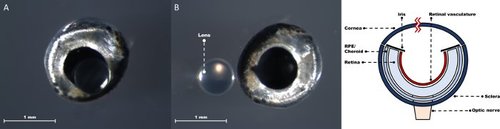
Adult zebrafish eye in the process of lens removal. Corneal side view with the lens pushed through the corneal tear (A). Corneal side view with the lens beside the ocular globe (B). Schematic depiction of the zebrafish eye at this step (C). |

Corneosclera consisting of sclera and cornea, which is disconnected from the remaining intraocular tissue. Corneal side view showing the continuation of the translucent cornea into the pigmented sclera (A). Lateral view focused on the scleral part of the corneosclera (B). Schematic depiction of the removed corneosclera (C). |
Adult zebrafish eye after removal of the corneosclera. Lateral view shows the remaining intraocular tissue with intact iris and optic nerve (A). Corneal side view with iris in focus (B). Schematic depiction of the zebrafish eye at this step (C). |

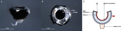
Adult zebrafish eye after removal of RPE/choroid and truncated optic nerve. Optic nerve side view shows the retina with a truncated optic nerve (A). Corneal side view with a direct look onto the most inner layer of the retina (B). Schematic depiction of the zebrafish eye at this step (C). |
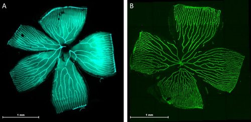
Representative pictures of adult tg(fli:EGFP) zebrafish retinal vasculature. Two typical morphological examples of the retinal vasculature are shown: The central optic artery spreads into 5-7 main vessels, which then branch into a succession of arcades. All further vessels drain into the circumferential vein (CV) limiting the outer part of each petal. Visualization via fluorescence microscopy at 2.5x magnification (A). Visualization of the retinal vasculature by confocal laser scanning microscopy through a combined 5x5 single image tile scan (B). |
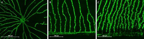
Magnified areas of adult tg(fli:EGFP) zebrafish retinal vasculature visualization. The optic artery branching into main vessels (A). Retinal capillaries in an area of low vascular activity connecting to the inner optic circle (IOC) at the bottom of the picture (B). Retinal capillaries in an area of high vascular activity connecting to the IOC (C). |

HE staining of the adult zebrafish eye. Cross-sectional overview of the whole eye after removal of the lens (A). Retinal layers of the adult zebrafish eye19 (B). Indication (black arrow) of the natural breaking point in the PL (C). |

Example presentation of vascular parameters as readout of retinal vasculature visualization. Measurement of intervascular distance (red double arrow) near the inner optic circle (IOC) (A). Three branching points (red circles) near the IOC (B). Visualization of zebrafish retinal vasculature with an enlarged area (red box) showing a sprouting vessel (C). Vessel diameter measured over a certain distance (white double arrow) from the central artery (white cross) (D). Vessel density is the percentage of retinal area occupied by overlaying vessels (diagonal red lines) (E). |

Comparison of HE staining and autofluorescence in the adult zebrafish retina. Retinal vasculature in focus above the GCL (A). Retinal vasculature (white box) showing green an EGFP signal and retinal layers exhibiting strong autofluorescence (B). |