
- Title
-
DNAAF1 links heart laterality with the AAA+ ATPase RUVBL1 and ciliary intraflagellar transport
- Authors
- Hartill, V.L., van de Hoek, G., Patel, M.P., Little, R., Watson, C.M., Berry, I.R., Shoemark, A., Abdelmottaleb, D., Parkes, E., Bacchelli, C., Szymanska, K., Knoers, N.V., Scambler, P.J., Ueffing, M., Boldt, K., Yates, R., Winyard, P.J., Adler, B., Moya, E., Hattingh, L., Shenoy, A., Hogg, C., Sheridan, E., Roepman, R., Norris, D., Mitchison, H.M., Giles, R.H., Johnson, C.A.
- Source
- Full text @ Hum. Mol. Genet.

Genetic complementation of heart looping defects in dnaaf1−/− mutant zebrafish embryos: Zebrafish embryos from dnaaf1+/− heterozygote-heterozygote inter-crosses were either mock injected (control, n = 405 embryos), or injected with mRNA expressed from human wild-type (wt, n = 425 embryos) and p.Leu191Phe-mutant (mut, n = 547 embryos) pCS2+ DNAAF1 constructs. In all cases, a mixture of normal looping (green), reversed looping (red) or ‘linear’ looping indicating a lack of heart looping (orange) were seen and representative examples are shown at 72 h post fertilization (left panels). On the right, the bar graph quantifies the three positions of heart looping in all three experiments, showing that phenotypic rescue is seen after injection of wild-type but not mutant RNA. n.s.= not significant; *** P < 0.001 (one-way ANOVA, n = 3 biological replicates). The dashed line indicates the 25% expected Mendelian ratio of embryos that should be homozygous mutant and manifest a phenotype. PHENOTYPE:
|
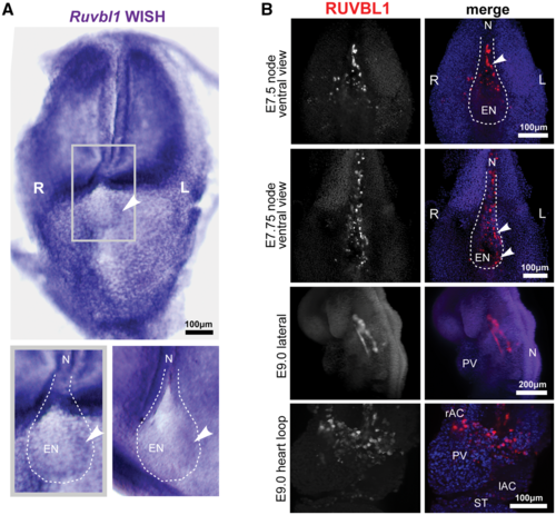
Left-sided asymmetric expression of RUVBL1 at the mouse embryonic node: (A) whole-mount in situ hybridization (WISH) of Ruvbl1 probes for whole-mount head-fold stage wild-type C3H/HeH mouse embryos showing widespread, ubiquitous Ruvbl1 expression and left-sided asymmetric distribution at the embryonic node. Grey frame indicates magnified inset (lower left) with a second example at lower right, with Ruvbl1 expression indicated by arrowheads. Scale bar = 100 μm. (B) Light-sheet microscopy images of whole-mount immunofluorescence for RUVBL1 (red) and DAPI (blue) in the wild-type C57BL/6 mouse E7.25 and E7.5 (late head fold and 2–4 somite stage) embryonic node, and E9.0 (6–8 somite stage) vasculature and heart loop. Left (L) and right (R) sides are indicated. RUVBL1 is asymmetrically distributed at the murine embryonic node (upper two panels). RUVBL1 is present in a widespread distribution throughout the atrial chambers, primitive ventricle and developing vasculature (lower two panels). Scale bars = 100 or 200 μm, as indicated. Abbreviations: rAC and IAC, right and left atrial chambers; EN, embryonic node; L, left; N, notochord; PV, primitive ventricle; R, right; ST, septum transversum. |
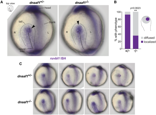
DNAAF1-dependent left-sided asymmetric expression of RUVBL1 at the zebrafish Kupffer’s vesicle: (A) in situ hybridization (ISH) of Ruvbl1 expression at Kupffer’s vesicle (thin dashed black line) in wild-type heterozygous dnaaf1+/− and mutant homozygous dnaaf1−/− zebrafish embryos, at the 5 to 9 somite stages, visualized from dorsal views as indicated in the top left inset. Localized expression is indicated by arrowheads, and the mid-line of the notochord is shown by the thick dashed black line. Left (L) and right (R) sides are indicated. (B) Bar graph shows a significantly higher proportion of wild-type zebrafish (+/−) have left-sided localisation of ruvbl1 when compared to diffused central localization in mutant (−/−) embryos (P = 0.0023, unpaired Student’s t-test). (C) Additional examples of ruvbl1 expression at Kupffer’s vesicle in unaffected dnaaf1+/− heterozygous and dnaaf1−/− affected mutant zebrafish embryos. EXPRESSION / LABELING:
PHENOTYPE:
|