
- Title
-
A Three-Layer Network Model of Direction Selective Circuits in the Optic Tectum
- Authors
- Abbas, F., Triplett, M.A., Goodhill, G.J., Meyer, M.P.
- Source
- Full text @ Front. Neural Circuits

Functional characterization of DS SINs. (A) Diagram showing dorsal view of the retinotectal projection in zebrafish larvae. Retinal ganglion cells (in yellow) send projections contralaterally from the retina to the neuropil of the optic tectum where they arborize. Periventricular neurons (PVNs, in pink) project dendrites into the tectal neuropil. Unlike PVNs, Superficial inhibitory interneurons (SINs) (cyan) have cell bodies located in the most superficial tectal neuropil and extend broad monostratified arbors into the retinorecipient layers. (B) Responses of a single SIN expressing GCaMP5G to a drifting grating stimulus. Red arrows indicate direction of grating motion, yellow arrow indicates SIN cell body and blue arrow indicates SIN arbor. White dashed line indicates skin covering the tectum. (C) Example response of a single SIN tuned to 140° directed motion. Directions of motion eliciting significant responses are indicated by arrows. Inset shows response as a polar plot. (D) Cumulative histogram of preferred directions of all DS SINs. Fitting von-Mises curves (red lines, R2 = 0.8) to the population histogram reveals three normally distributed, non-overlapping populations with population peaks centered at 9°, 157°, and 264°. Each population is color-coded according to preferred direction. (E) Normalized responses of direction selective SINs, color-coded according to subtype. Responses of individual cells are shown as colored lines, mean responses shown in black. (F) Polar plots showing normalized responses of each population; mean (solid line) and dashed (±1 SD). Mean DSI, tuning bandwidth (full width half max) and preferred direction of each population relative to the body axis of the fish are shown below each polar plot. |
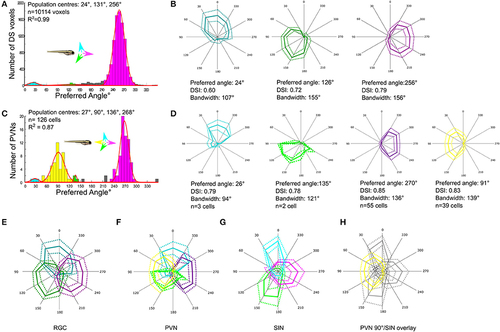
Comparison of direction-selectivity in the SIN, RGC and periventricular neuron (PVN) populations. (A) Cumulative histogram of preferred directions of all RGC voxels (n = 10,114 voxels from 29 larvae). Fitting von-Mises curves (red lines, R2 = 0.99) to the population histogram reveals three normally-distributed, non-overlapping populations with population peaks centered at of 24°, 131°, and 256°. Each population is color-coded and the preferred direction of motion of each population relative to the body axis of the fish is shown in inset. (B) Polar plots showing normalized responses of each DS RGC subtype; mean (solid line) and dashed (±1 SD). Mean DSI, tuning bandwidth (full width half max) and preferred direction of each DS RGC subtype are shown below each polar plot. (C) Histogram of DS PVN preferred directions (n = 126 tectal neurons from 24 fish). Four normally distributed populations are present, with populations centered at 27°, 90°, 136°, and 268° (R2 = 0.87). Note the emergent population of PVN tuned to 90° motion (yellow) that is absent from both RGCs and SINs. (D) Polar plots showing normalized responses of each DS PVN subtype; mean (solid line) and dashed (±1 SD). Mean DSI, tuning bandwidth (full width half max) and preferred direction of each subtype are shown below each polar plot. (E–H) Overlaid polar plots summarizing how direction of motion is encoded by RGCs (E), PVNs (F), and SINs (G). RGCs exhibit a tiled and triangular representation of directional space by three subtypes of DS RGC. In the SIN population the representation of motion is reshaped, leaving a gap at 90°. Directional space is represented by 4 subtypes of PVNs with preferred directions aligned with the cardinal axes. (H) Overlaying the polar plots of DS SINs with the emergent population of PVN reveals that the gap in the representation of motion in the rostral-to-caudal direction in the SIN population aligns with the emergent population of PVNs tuned to 90°. |