
- Title
-
Evaluation of (fli:GFP) Casper Zebrafish Embryos as a Model for Human Conjunctival Melanoma
- Authors
- Pontes, K.C.S., Groenewoud, A., Cao, J., Ataide, L.M.S., Snaar-Jagalska, E., Jager, M.J.
- Source
- Full text @ Invest. Ophthalmol. Vis. Sci.
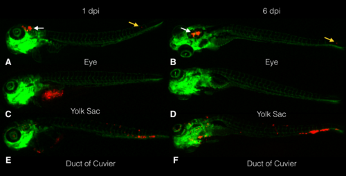
Stereo fluorescence image of zebrafish embryos engrafted with CM cells (vasculature in green and CM cells in red). The embryos were injected at 2 dpf with CRMM-1 CM cells labeled with tomato-red (red). Photographs taken of the same embryo that had been injected with CM cells around the eye at 1 (A) and 6 dpi (B), showing cells inside the head (white arrows) and in the tail (yellow arrow). Following injection in the yolk sac, an embryo shows the cells in the yolk sac at 1 (C), but not 6 dpi (D). After injection of cells into the duct of Cuvier, cells are seen inside the circulation at 1 dpi (E), mainly in the tail and inside the eye. The same embryo shows a cluster in the tail and cells inside the eye at 6 dpi (F). The stereo fluorescent images (original magnification: ×20) are representative of >10 independent experiments. EXPRESSION / LABELING:
|
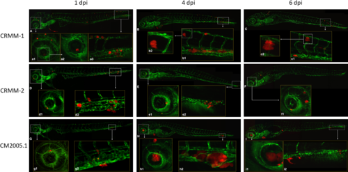
Confocal micrographs of the observed phenotypes at 1, 4, and 6 dpi after engraftment of three CM cell lines via the duct of Cuvier in (fli:GFP) Casper zebrafish embryos. At 1 dpi, CRMM-1 (A), CRMM-2 (D), and CM2005.1 (G) cells were already inside the eye (a1, a2, d1, g1) and in the tail (a3, d2, g2). At 4 (B, E, H), and 6 dpi (C, F, I), cells formed clusters in the tail and in the eye in all three cell lines (data not shown). The clusters were more evident in the tail (h2) and in the eye (h1, i1) after injection of cell line CM2005.1. The three cell lines (data not show) grew inside (a3, b1, d2, e2, g2, h2, i2), outside (b2), and around (c2) the vessels and the cells could be found inside the eye (f1, i1) until 6 dpi. The images were acquired using a Leica TCS SPE confocal microscope and managed in ImageJ software. Images (A–I) ×10 dry objective. All the other images: ×20 dry objective. Red: cells labeled with tdTomato; green: GFP-endothelial cells of the (fli:GFP) Casper lines. EXPRESSION / LABELING:
|
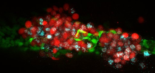
Confocal image of immunohistochemistry with Ki67 in a whole 6 dpi (fli:GFP) Casper zebrafish embryo. There were 200 to 400 CRMM-1 td-Tomato CM cells injected into the duct of Cuvier. We see tumor cell (red) migration outside the vessels (green); cell proliferation is indicated by Ki67 staining (blue). This image of the tail of a live embryo was acquired by confocal microscope (×20 dry objective). Similar images were obtained from all three CM cell lines in >10 independent experiments. |