
- Title
-
Neurotrophin-conjugated nanoparticles prevent retina damage induced by oxidative stress
- Authors
- Giannaccini, M., Usai, A., Chiellini, F., Guadagni, V., Andreazzoli, M., Ori, M., Pasqualetti, M., Dente, L., Raffa, V.
- Source
- Full text @ Cell. Mol. Life Sci.

5 dpf larvae injected with 2 nl of various doses of H2O2 and fixed 8 hpi. a–d TUNEL and Hoechst staining: representative sections of larvae injected with 0 (k), 0.25 M, 0.5 M or 1 M H2O2. e Time course of the experiment. f Quantitative analysis of TUNEL-positive cells in Ph, INL, GCL and CMZ cell layers for the different doses. n ≥ 15 (embryos), 1-way ANOVA followed by Bonferroni correction (all groups compared against k group). Ph: p = 0.04. INL: p < 0.0001. GCL: p < 0.0001. CMZ: p = 0.0003 |

Larvae (4 dpf) were injected with the nanoparticles, and localization was studied 24 hpi (a). Representative images of particles localized in RPE (B1) or in the NR (B2). Particles never localise outside the ocular tissues (B3). Particles are stained blue (Prussian blue). The bars are 100 µm. c Normalized distribution of MNP staining in the NR, RPE and the choroidal layer. n > 15, 2-way ANOVA, p = 0.27 |

NGF was labeled with Alexa Fluor 488 (NGFfluo). MNP–NGFfluo were injected into larvae (4 dpf) and the localization of MNPs and NGFfluo was studied 6 hpi. MNPs (brown signal shown by red arrows in B1) and NGFfluo (green staining in B2) were found to co-localise in the GCL |

Retina, RPE and choroid are shown in an eye section of 5 dpf larva. |
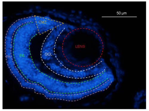
The boundaries of lens (red dotted lined), GCL (white dotted lines), INL (green dotted lines), Ph (pink dotted lines) and CMZ (yellow dotted lines) are schematically represented in an eye section of 5 dpf larva. |

Representative images of 5 dpf larvae injected with 2 nl of 1 M H2O2 and fixed 2 or 4 or 8 or 24 hours post injection (A, B, C and D, respectively): TUNEL staining (1) and Hoechst staining (2) and their merge (3). |