
- Title
-
Therapy-induced developmental reprogramming of prostate cancer cells and acquired therapy resistance
- Authors
- Nouri, M., Caradec, J., Lubik, A.A., Li, N., Hollier, B.G., Takhar, M., Altimirano-Dimas, M., Chen, M., Roshan-Moniri, M., Butler, M., Lehman, M., Bishop, J., Truong, S., Huang, S.C., Cochrane, D., Cox, M., Collins, C., Gleave, M., Erho, N., Alshalafa, M., Davicioni, E., Nelson, C., Gregory-Evans, S., Karnes, R.J., Jenkins, R.B., Klein, E.A., Buttyan, R.
- Source
- Full text @ Oncotarget
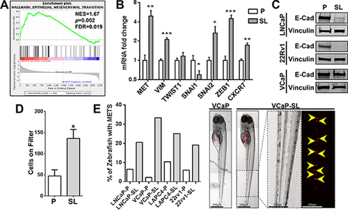
Reprogrammed PCa cells possess increased invasive/metastatic abilities. (A) GSEA indicates a significant enrichment of EMT genes in LNCaP-SL cells. (B) qPCR analyses show that reprogrammed LNCaP-SL cells overexpress mRNA of key EMT markers. Mean ± standard error; *P < 0.05; **P < 0.01; ***P < 0.001. (C) Western Blot demonstrates loss of E-Cadherin in reprogrammed PCa cells indicating increased EMT phenotype. (D) Reprogrammed LNCaP-SL cells demonstrate increased invasion through a Matrigel-coated membrane in in vitro invasion assays compared to parental LNCaP cells. Mean ± standard error; *P < 0.05. (E) Reprogrammed stem-like PCa cells are more invasive/metastatic in zebrafish assays (Left). Representative photomicrographs of VCaP and VCaP-SL xenografted fish illustrates dispersal of fluorescently-labelled VCaP-SL cells within the caudal region (Right). |