
- Title
-
Acceleration of osteoblast differentiation by a novel osteogenic compound, DMP-PYT, through activation of both the BMP and Wnt pathways
- Authors
- Bae, S.J., Kim, H.J., Won, H.Y., Min, Y.K., Hwang, E.S.
- Source
- Full text @ Sci. Rep.
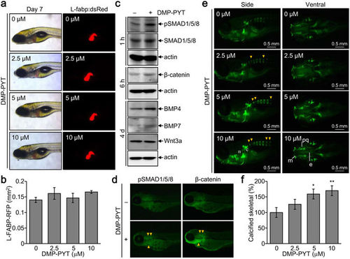
Increased calcified skeletal development by DMP-PYT in vivo. L-Fabp:dsRed zebrafish embryos (n = 10) were incubated with the indicated amounts of DMP-PYT for 6–7 days post fertilization. (a) Zebrafish were observed under a fluorescence stereomicroscope and ref fluorescence was observed in the liver. (b) The hepatic size showing red fluorescence was quantitatively determined from 10 larvae images by automatically calculating the fluorescent area (mm2) in the liver. (c,d) Zebrafish larvae (n = 10) at day 3 post fertilization were treated with DMP-PYT for 1 h, 6 h, and 4 days and harvested for immunoblotting analysis. Protein blots were incubated with antibodies against pSMAD1/5/8, SMAD1/5/8, β-catenin, BMP4, BMP7, Wnt3a, β-actin, followed by enhanced chemiluminescence and autoradiography (c). Fish larvae that were treated with DMP-PYT for 1 and 6 h and subjected to immunohistochemistry of pSMAD1/5/8 and β-catenin (d). (e) Skeletal development of zebrafish was visualized by calcein staining at day 7 post fertilization. Side view of the head skeleton: n, notochord; c, cleitrum. Ventral view of the head skeleton: m, Meckel’s cartilage; pq, palate quadrate; e, ethmoid plate. Arrowheads indicate the increased bone formation in vertebrate skeleton. (f) Calcification of axial skeleton was quantitatively determined from 10 larvae images using image J software and expressed as the percentage of the control. *P < 0.05 and **P < 0.005. EXPRESSION / LABELING:
PHENOTYPE:
|