
- Title
-
Identification and characterization of variants and a novel 4 bp deletion in the regulatory region of SIX6, a risk factor for primary open-angle glaucoma.
- Authors
- Shah, M.H., Tabanera, N., Krishnadas, S.R., Pillai, M.R., Bovolenta, P., Sundaresan, P.
- Source
- Full text @ Mol Genet Genomic Med
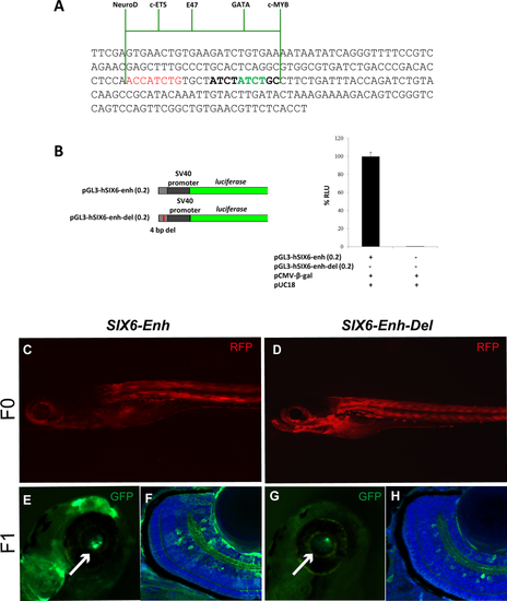
Functional characterization of the 4 bp deletion in the SIX6 enhancer found in POAG patients. (A) Sequence of the short enhancer fragment used to perform luciferase assays. The deleted sequence is indicated in green, the NeuroD-binding site in red. The position of binding sites for the transcription factors ETS, E47, GATA, and C-MYB is indicated with green lines. (B) Schematic representation of the constructs used to perform luciferase assays (left) and graph of the luciferase assay performed with the two different constructs and control plasmids as indicated in the panel. Bars represent % mean values ± SEM normalized to control; ***P < 0.0001. (C–H) Images of representative transgenic embryos generated with a human reference SIX6 retinal enhancer (C, E, F) and an enhancer containing the 4 bp deletion found in POAG patients (D, G, H). Embryos in (C, D) represent F0 transgenic embryos expressing control RFP at similar levels. (E–H) Images of representative F1 transgenic embryos showing GFP reporter expression driven by the reference (E, F) or deleted enhancer (G, H). Images in (e, g) are high power in toto view of the eye (arrows), whereas those in (F, H) are the corresponding frontal cryostat sections. Note how the reference enhancer drives strong GFP expression (green) in cells of the retinal ganglion and amacrine layers (F), whereas the deletion strongly downregulates reporter expression (H). Sections are counterstained with Hoechst (blue). Abbreviations: acl, amacrine cell layer; gcl, ganglion cell layer. EXPRESSION / LABELING:
|