
- Title
-
Mitochondrion to endoplasmic reticulum apposition length in zebrafish embryo spinal progenitors is unchanged in response to perturbations associated with Alzheimer's disease
- Authors
- Newman, M., Halter, L., Lim, A., Lardelli, M.
- Source
- Full text @ PLoS One

The midline of the spinal cord region. M-ER apposition lengths were measured in 3–6 cells at the midline area of the spinal cord at each embryo (such as those indicated by white arrowheads). Three embryos were examined for each treatment. The white, dashed line indicates the midline of the spinal cord. PHENOTYPE:
|
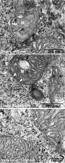
Electron microscopy of zebrafish neural cells. Cells were treated with morpholinos binding the start codon of psen1 and psen2 mRNA (MoPS1Tln and MoPS2Tln respectively). Arrowheads indicate the region of apposition between mitochondria (M) and endoplasmic reticulum (E) i.e. MAM. PHENOTYPE:
|

Unillustrated author statements |