
- Title
-
Characterization of trace metal content in the developing zebrafish embryo
- Authors
- Thomason, R.T., Pettiglio, M.A., Herrera, C., Kao, C., Gitlin, J.D., Bartnikas, T.B.
- Source
- Full text @ PLoS One
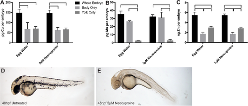
Distribution of metals in bodies and yolk sacs of embryos. (A-C) ICP-MS measurements were collected for copper (Cu) (A), manganese (Mn) (B), and zinc (Zn) (C) in whole embryos, bodies only, and yolk sacs only of embryos at 48 hpf, reared in presence or absence of 5 μM neocuproine, a Cu chelator. Each bar represents the average and standard deviation of three independently collected replicates, with each replicate consisting of a pooled group of 47–50 embryos. Horizontal brackets above bars indicate statistically significant difference between bracketed values. (Data in all panels passed Shapiro-Wilk normality and equal variance tests. One-way ANOVA indicated significant difference between groups, with the Holm-Sidak method used for multiple comparisons and P<0.05 as cut-off for significance.) There was no significant difference in metal levels between neocuproine-treated and–untreated samples of the same sample type. (D, E) Images were taken of zebrafish embryos at 48 hpf, reared in presence or absence of 5 μM neocuproine. |