
- Title
-
A small molecule screen identifies in vivo modulators of peripheral nerve regeneration in zebrafish
- Authors
- Bremer, J., Skinner, J., Granato, M.
- Source
- Full text @ PLoS One

Pectoral fin innervation and nerve regrowth after fin removal. (A-B) Dorsal (A) and side view (B) of a larval zebrafish at 5 dpf showing the location of the pectoral fin (magenta dashed box). (C, C') Pectoral fin innervation in a Tg(mnx1:GFP) transgenic larva at 5 dpf (C) and a schematic (C'). The first segmental nerve (orange) forms the dorsal plexus together with the joint nerves 2 and 3 (red). The fourth nerve (green) travels along the larval body wall, before it enters the pectoral fin ventrally. These nerves contribute to the ring-like network at the fin base (blue). (D-E) Fin region of Tg(mnx1:GFP) transgenic larvae 1 hour (D) and 24 hours (E) after fin removal. One hour after the pectoral fin has been removed using tweezers, GFP positive axons at the fin base are no longer detectable, leaving behind the stumps of nerve 1–3 (orange, red) at the dorsal plexus and at the 4th nerve entry point (green, D). Twenty-four hours after fin removal, GFP positive axons at the fin base have regrown robustly, reforming the ring-like nerve network at the fin base (blue) in 100% of the 66 control fish tested (untreated and DMSO treated, E). |

Nerve regrowth at the fin base requires sox10 and FGF. (A-C) Quantification of fin nerve regrowth in Tg(mnx1:GFP) transgenic larvae 24 hours after fin removal; nerves show different degrees of nerve regrowth (A-C). Categories 0, 1, and 2 indicate that none (A, A'), one (B, B'), or both sides (C,C') of the ring-like nerve network at the fin base regrew, respectively. (A'-C'): schematic representation of these results. (D-F) Nerve regrowth at the fin base 24 hours after fin removal in a wild type sibling (D) and a sox10 mutant (E). Sox10 mutants display significantly reduced nerve regrowth compared to wild type siblings. Graphical representation of the extent of nerve regrowth (category 0, 1, 2; F). (G-J) Nerve regrowth at the fin base at 24 hours after fin removal in a control larva treated with DMSO (G) and larvae treated with a low (17μM, H) or a high dose (34μM, I) of the FGF inhibitor SU5402. DMSO or SU5402 were added immediately after fin amputation. FGF inhibitor-treated larvae show significantly less nerve regrowth compared to DMSO-treated controls. Graphical representation of the extent of nerve regrowth (category 0, 1, 2; J). All scale bars are 10 μm. |

FGF inhibitor and six additional compounds that impair nerve regeneration after fin removal also impair regeneration after laser nerve transection. (A) Ventral nerves of Tg(mnx1:GFP) transgenic larvae were laser transected and treated with DMSO for control, or treated with a low (17μM) or high (34μM) dose of the FGF inhibitor SU5402. Regeneration was scored 48 hours later and the graphical representation of the extent of nerve regeneration (no/ weak, moderate, strong regeneration) is shown, demonstrating that the FGF inhibitor SU5402 significantly and dose-dependently impairs nerve regeneration. (B) Quantification of dorsal nerve regeneration in Tg(isl1:GFP) transgenic larvae measuring the extent of dorsal nerve regeneration (no/ weak, moderate, strong regeneration) for controls (0.5% DMSO) and all tested compounds. Post nerve transection exposure to 100μM anandamide (cannabinoid agonist), 25μg/ml HA-1004 (PKC inhibitor), 10μg/ml phenamil (TRPP3 channel blocker), and 25μg/ml MnTBAP (SOD mimetic) did not significantly impair nerve regeneration. However, 25μg/ml lavendustin (EGFR inhibitor), 4μM AM-580 (RA receptor agonist), 10μg/ml verapamil (calcium channel blocker), 5μM PGD2, 25μg/ml dexamethasone (corticosteroid), and 250μM 10-HCT significantly impaired nerve regeneration. (C-I) Ventral nerves of Tg(mnx1:GFP) transgenic larvae treated with DMSO for control (C, D), or with 125μM of the topoisomerase I inhibitor 10-HCT (E-H) before laser nerve transection (C, E, G) and 48 hours later (D, F, H). Control DMSO-treated larva showing normal morphology of the ventral nerve before transection (C), and robust nerve regeneration after 48 hours (D). Transected nerves treated with 10-HCT showed normal morphology before transection (E), but failed to regenerate after injury (F). In contrast, an untransected ventral nerve treated with 10-HCT showed normal morphology at all times (G,H). Graphical representation of the results, showing that 10-HCT significantly impaired nerve regeneration (I). (J) Change in the number of GFP-labeled neuronal cell bodies in Tg(isl1:GFP) transgenic larvae between day 5 and day 7, treated with DMSO for control or with 125μM 10-HCT. Dorsal nerves were either left untransected or laser transected at day 5. On average, 26 neurons were labeled per segment. There was no difference in the change of the neuronal cell body number between DMSO and 10-HCT treated larvae when left untransected or after nerve transection, suggesting that 10-HCT did not reduce neuronal survival. However, following laser nerve transection in both DMSO and 10-HCT treated larvae, the number of neurons decreased on average by 3 neurons, suggesting that a subset of neurons with transected axons died. |
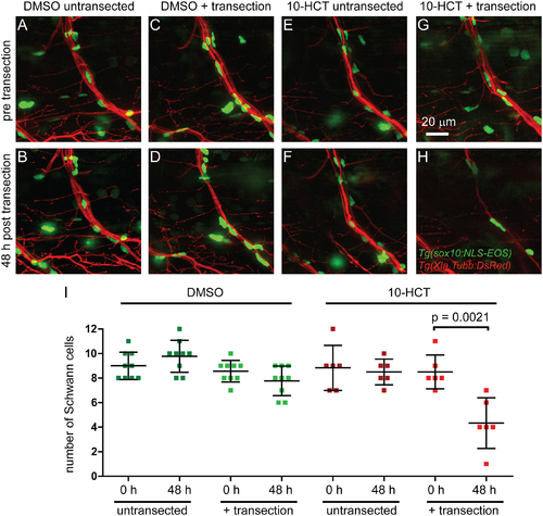
Topoisomerase I is required for survival of denervated Schwann cells following laser nerve transection. Ventral nerves of Tg(Xla.Tubb:DsRed), Tg(sox10:NLS-Eos) double transgenic larvae treated with DMSO for control (A-D), or with the topoisomerase inhibitor 10-HCT (E-H); untransected controls (A,B,E,F) or laser nerve transected (C,D,G,H) at different time points: before transection (A,C,E,G) or 48 hours post transection (B,D,F,H). Graphical representation of the number of Schwann cells for the different conditions and time points (I). In DMSO controls, the number of Schwann cells was only non-significantly marginally reduced after laser nerve transection (C,D). Similarly, in untransected control nerves, 10-HCT did not reduce the number of Schwann cells, excluding the possibility that 10-HCT is simply toxic to Schwann cells. However, after laser nerve transection, the number of Schwann cells was significantly reduced, suggesting that 10-HCT specifically ablated denervated Schwann cells. |