
- Title
-
Augmented quantal release of acetylcholine at the vertebrate neuromuscular junction following tdp-43 depletion
- Authors
- Dzieciolowska, S., Drapeau, P., Armstrong, G.A.B.
- Source
- Full text @ PLoS One
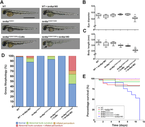
tardbpY220X/Y220X zebrafish embryos injected with an ATG tardbpl morpholino (MO) displayed significant morphological defects and reduced survival. A, Representative images of 2 dpf larvae from the following treatment groups: WT, WT + MO, tardbpY220X/+, tardbpY220X/Y220X, tardbpY220X/Y220X + Control MO (CoMo), tardbpY220X/Y220X + MO. Scale bar represents 1 mm, arrow indicates cardiac defect in the tardbpY220X/Y220X + MO treated larva. B, Eye diameter and C, body length was determined for each treatment group. tardbpY220X/Y220X + MO 2dpf larvae displayed significantly reduced eye diameter compared to all other treatment groups (p < 0.01). WT + MO, tardbpY220X/Y220X and tardbpY220X/Y220X + CoMo larvae displayed significantly reduced body length compared to WT larvae (p < 0.05) and tardbpY220X/Y220X + MO displayed significantly reduced body length compared to all treatment groups (p < 0.01). D, Quantification of gross morphological defects observed in all treatment groups indicating a higher incidence of defects in the tardbpY220X/Y220X + MO condition. E, Percentage survival curves of all treatment groups tracked over 11 days. tardbpY220X/Y220X + MO-treated larvae displayed reduced survival, with all animals dying by 10 dpf (p < 0.0001). N = 2, n = 50, *p < 0.05; **p < 0.01. |

ZFIN is incorporating published figure images and captions as part of an ongoing project. Figures from some publications have not yet been curated, or are not available for display because of copyright restrictions. PHENOTYPE:
|
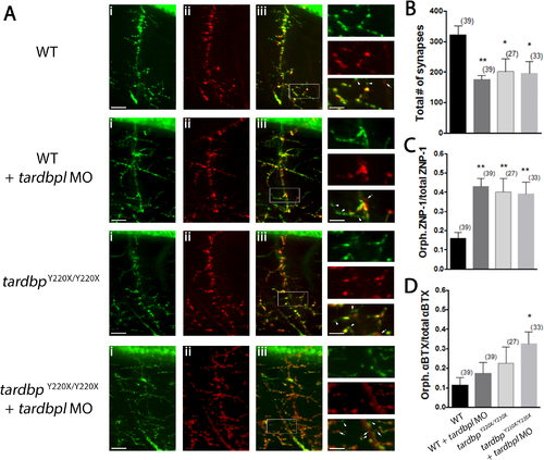
Motor neuron projections in tardbpY220X/Y220X + MO larvae show increased number of orphaned presynaptic and postsynaptic puncta. A, Representative images of single ventral root projection double-labelled for ZNP-1 (presynaptic marker, i) and sulforhodamine-conjugated αBTX (postsynaptic marker, ii). WT larvae display extensive co-localization of both ZNP-1 and αBTX (merged image, iii). Scale bar in (iii) and insets represent 25 μm and 10 μm respectively, arrowheads indicate orphaned ZNP-1 puncta and arrows indicate orphaned αBTX labelling. B, Quantification of the number synapses formed, quantified as the number of colocalized ZNP-1 and αBTX puncta. WT + MO (p < 0.05), tardbpY220X/Y220X (p < 0.01) and tardbpY220X/Y220X + MO larvae (p < 0.01) all had a significantly reduced number of synapses at the NMJ compared to WT larvae. C, Quantification of orphaned presynaptic ZNP-1 puncta over total number of ZNP-1 puncta. WT + MO, tardbpY220X/Y220X and tardbpY220X/Y220X + MO larvae displayed significantly higher proportion of orphaned ZNP-1 puncta compared to WT animals (p < 0.01). D, Quantification of orphaned postsynaptic αBTX staining over total number of αBTX puncta. tardbpY220X/Y220X + MO fish displayed significantly higher proportion of orphaned αBTX puncta compared to WT larvae (p < 0.05). Numbers in parentheses represent the number of somites analyzed for each treatment group. Data expressed as mean ± SEM: *p < 0.05; **p < 0.01. PHENOTYPE:
|