
- Title
-
AUTEN-67, an autophagy-enhancing drug candidate with potent antiaging and neuroprotective effects
- Authors
- Papp, D., Kovács, T., Billes, V., Varga, M., Tarnóci, A., Hackler, L., Puskás, L.G., Liliom, H., Tárnok, K., Schlett, K., Borsy, A., Pádár, Z., Kovács, A.L., Hegedűs, K., Juhász, G., Komlós, M., Erdős, A., Gulyás, B., Vellai, T.
- Source
- Full text @ Autophagy
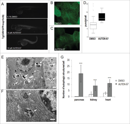
AUTEN-67 enhances autophagy in zebrafish and mice. (A) AUTEN-67 increases the glowing intensity of an autophagy reporter (Gfp-Lc3b) in zebrafish embryos. GFP intensity increases by 1.36- and 1.38-fold upon administration of 10 and 50 µM of AUTEN-67, respectively (P < 0.001, paired Student t test). Fluorescent images. N=4, at both concentrations. (B) Fluorescence image of an untreated (DMSO) Gfp-Lc3 fish sample (large magnification). (C) Fluorescence image of a sample treated with AUTEN-67. Green foci indicate autophagic structures. (D) Quantification of green punctae in untreated vs. treated cells. P < 0.01; paired Student t test. (E) Transmission electron microscopy (TEM) image showing the ultrastructure of exocrine pancreatic tissue from a mouse treated with AUTEN-67. Arrows indicate autophagic structures (autophagosomes and autolysosomes). Note that control cells display almost no sign of autophagic structures observed at the ultrastructural level.47 Kovács J, László L, Kovács AL. Regression of autophagic vacuoles in pancreatic acinar, seminal vesicle epithelial, and liver parenchymal cells: a comparative morphometric study of the effect of vinblastine and leupeptin followed by cycloheximide treatment. Exp Cell Res 1988; 174:244-51; PMID:3335225; http://dx.doi.org/10.1016/0014-4827(88)90158-9 [Google Scholar] Scale bar: 1 µm. (F) TEM picture displays the ultrastructure of a pancreatic cell from a mouse administered orally with AUTEN-67. Arrows indicate late autolysosomes, implying that AUTEN-67 induces autophagic degradation. Scale bar: 1 µm. (G) Quantification of autophagic structures in exocrine pancreatic (“pancreas”), kidney epithelial (“kidney”) and heart muscle (“heart”) tissues from mice treated with AUTEN-67 solved in DMSO (red columns) and with DMSO only (blue columns). Bars represent s.e. In each tissue examined, differences between the corresponding treated and untreated samples are statistically significant (***: P < 0.001; paired Student t test). |