
- Title
-
Gpr161 anchoring of PKA consolidates GPCR and cAMP signaling
- Authors
- Bachmann, V.A., Mayrhofer, J.E., Ilouz, R., Tschaikner, P., Raffeiner, P., Röck, R., Courcelles, M., Apelt, F., Lu, T.W., Baillie, G.S., Thibault, P., Aanstad, P., Stelzl, U., Taylor, S.S., Stefan, E.
- Source
- Full text @ Proc. Natl. Acad. Sci. USA
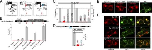
Cellular PPIs and localization of Gpr161 variants. (A) Schematic illustration of the Rluc-PCA biosensor strategy to quantify PPIs of wild-type and mutated Gpr161 and RI in vivo. Mutated domains are highlighted in red/blue. (B) Shown are conserved sequence elements in the Gpr161-CT. Impact of L465P mutation of Gpr161-F[1]/[2] on complex formation with RIα-F[1]/[2] (±SEM; representative of n = 3 independent experiments; murine Gpr1611-528; NP_001297359.1). (C) Impact of Gpr161 mutations of the flanking Leu of the PPI-motif and the PKA phosphorylation consensus site on RIα:Gpr161 PPI. Read out: Rluc PCA (±SEM of at least n = 4 independent experiments). (D) Impact of the L50R mutation on RIβ:Gpr161 PPI; Rluc PCA measurements (±SEM; representative of n = 3). (E) Subcellular localization of mCherry-tagged Gpr161 or GFP-tagged RIα hybrid proteins in HEK293 cells. (Scale bar, 5 µm.) (F) Subcellular localization of coexpressed mCherry-tagged Gpr161 variants and GFP-tagged RIα in HEK293 cells. (Scale bar, 5 µm.) |
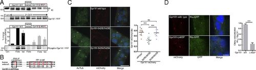
Phosphorylation and ciliary localization of Gpr161:RIα complexes. (A) IP of Venus-YFP-tagged Gpr161 variants expressed in HEK293 cells following treatments with Forskolin (20 µM, 10 min) and isoproterenol (1 µM, 10 min). Densitometric quantification of n = 4 independent experiments, ±SEM; phospho-(K/R)(K/R)X(S*/T*) specific antibody. The IB with the RI antibody is taken from a different experiment (better separation of antibody and RI). (B) Sequence comparison of AKAP and phosphorylation motifs from human and zebrafish Gpr161-CT. (C) Subcellular localization of indicated Gpr161-mCherry variants and acetylated-Tubulin in zebrafish. (Scale bar, 10 µm.) The graph shows the ratio of Gpr161-positive cilia and the total number of cilia in a minimum of three sections from three independent experiments. P values were calculated using one-way ANOVA and Tukey′s multiple-comparison post hoc test (***P < 0.001). Shown are the individual ratios (±SD) of Gpr161-mCherry wild-type (12 sections, 578 cilia), Gpr161-mCherry S428D/S429D (9 sections, 584 cilia), and Gpr161-mCherry S428A/S429A (9 sections, 415 cilia). (D) Coexpression of Gpr161-mCherry and RIα-GFP in zebrafish. The graph shows the ratio of RIα, Gpr161 double-positive cilia over the total number of Gpr161-mCherry-positive cilia (mean ± SEM, Gpr161-mCherry wild-type n = 7 embryos, Gpr161-mCherry L465P n = 6 embryos). ***P < 0.001 using two-tailed unpaired Student’s t test. (Scale bar, 5 µm.) |
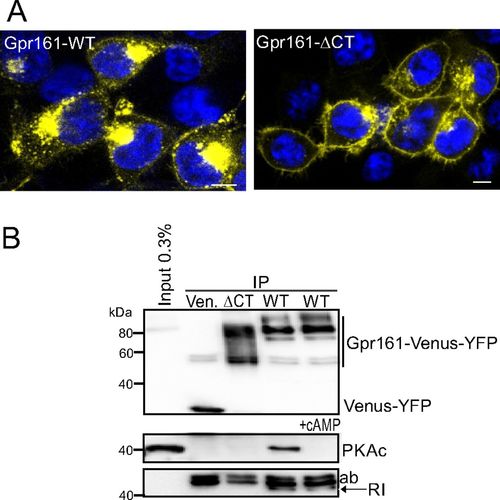
IP and localization of Gpr161. (A) Subcellular localization of Venus-YFP tagged Gpr161 fusion proteins in HEK293 cells. (Scale bars, 5 µm.) (B) IP of Venus-YFP tagged Gpr161 variants expressed in HEK293 cells (representative of n = 3 independent experiments). |