
- Title
-
Evolution and expression of the phosphodiesterase 6 genes unveils vertebrate novelty to control photosensitivity
- Authors
- Lagman, D., Franzén, I.E., Eggert, J., Larhammar, D., Abalo, X.M.
- Source
- Full text @ BMC Evol. Biol.
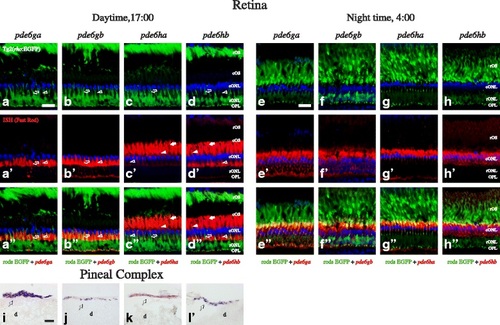
Expression pattern of the PDE6 inhibitory subunit genes in the adult zebrafish retina and pineal complex. Photomicrographs from ISH experiments on adult Tg2(rho:EGFP) zebrafish showing staining for pde6ga, pde6bg, pde6ha and pde6hb in the retina at 17:00 PM (a-d′′) and at 4:00 AM (e-h′′), and also in the pineal complex (i-l). The upper row shows EFGP fluorescence (green) in rods (a-h), the second row from the top shows the ISH experiments stained with Fast Red (red), (a′-h′), the second from the bottom is a merge of the two previous (a′′-h′′) and the bottom row shows ISH stained with NBT/BCIP (i-l). DAPI, in blue, stains cell nuclei in all fluorescent photomicrographs. Stricken morphological differences, due to the retinomotor movements, can be observed in the retina between day and night, graphically represented by the contraction of the rOS from the outermost layer at daytime (a-d) to inner locations at night (e-h). This makes very difficult the cell assignment in the night retinae, so it was shown only in the day retinae (a-d′′). Staining for pde6ga (a′-a′′ and e′-e′′) and pde6gb (b′-b′′ and f′-f′′) can be observed in rod inner segments and for pde6ha (c′-c′′ and g′-g′′) and pde6hb (d′-d′′ and h′-h′′) in cones. Staining for all four genes was observed in adult pineal complex (i-l). In a′-a′′ and b′-b′′, staining in the SSC seems to be present, therefore, we cannot discard that pde6ga and pde6gb are expressed in rods and SSC. In addition, the yellowish colour observed in 5g′′ could suggest double staining; however its absence in the day retina, combined with the about 100 fold lower expression levels and the loose morphology of the night retinae led us to conclude that this is just an artefact. Abbreviations: d; diencephalon, cONL; cone outer nuclear layer, cOS; cone outer segments, OPL; outer plexiform layer, rONL; rod outer nuclear layer, rOS; rod outer segments. Arrows point at DC, arrowheads to LSC, empty arrowheads to SSC and empty arrows to rods′ myoids. In i-l, arrows point at the pineal complex. Scale bars; in a is 20 µm for a-d′′, in e is 20 µm for e-h′′ and in i is 30 µm for i-l EXPRESSION / LABELING:
|
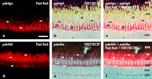
Coexpression in AB zebrafish retina of the 3R generated PDE6 inhibitory subunit paralogs. Photomicrographs from double ISH experiments on adult zebrafish outer retinae show colocalisation for pde6ga and pde6gb in rods (a-c) and for pde6ha and pde6hb in cones (d-f), arrows are pointing at the same cell in the three corresponding pictures. a and d are fluorescent pictures from Fast Red staining and b and e are bright-field pictures from, mainly, NBT/BCIP staining in purple but also a minimal staining from the Fast Red. c and f combine bright-field and fluorescence to show co-staining in the same cells. a-c shows exclusive staining in the myoids of the rods. The sections were obtained in an oblique angle to facilitate the perception of the rods´ myoids “crawling” between the cones, despite giving an unreal perception of the stratification. d-f show staining in the three cone types: SSC, LSC and DC. However, the tight packing of the DC makes it difficult to visualise them with fluorescence (d, f). Stars mark the cone-specific ellipsoids. Scale bar is 25 µm. RPE; retinal pigment epithelium. For the rest of abbreviations see Fig. 5 legend EXPRESSION / LABELING:
|

ZFIN is incorporating published figure images and captions as part of an ongoing project. Figures from some publications have not yet been curated, or are not available for display because of copyright restrictions. EXPRESSION / LABELING:
|

Retinomotor movements in the zebrafish retina. Photomicrographs of adult zebrafish retinae showing morphological differences between day (17:00; left column) and night (4:00; right column). In the upper row, bight field photomicrographs taken using Nomarski contrast show the clear stratification during daytime (a), different to the poorly stratified retina at night (b). In both pictures, Fast Red staining, in pink, shows the presence of pde6ga mRNAs in rods. Note the different intensity in staining, confirmed by qRT-PCR as higher expression at 4:00 AM (Fig. 7). c-f pictures are fluorescence photomicrographs of immunostainings illustrating the retinomotor movements with photoreceptor-specific markers. In c and d, sections of a transgenic line that expresses EGFP in cones: Tg(gnat2:EGFP) were incubated with a rod-specific anti-GNB1 antibody (in red). In e and f, sections of a transgenic line that expresses EGFP in rods: Tg2(rho:EGFP) were incubated with a double cone-specific anti-zpr1 antibody (in red). In both cases, note the position of the rod outer segments in the outermost part of the retina at daytime, while at night they have moved to inner positions and the cone outer segments have moved outwards. DAPI was used as a nuclei counterstain (c-f). RPE: retinal pigment epithelium. For more abbreviations, see Fig. 5 legend. Scale bar is 20 µm |