
- Title
-
Insights into kidney stem cell development and regeneration using zebrafish
- Authors
- Drummond, B.E., Wingert, R.A.
- Source
- Full text @ World J Stem Cells
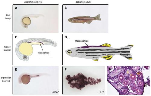
A, B: Live image of an embryonic zebrafish at 24 h post fertilization (hpf) and at the adult stage, respectively; C: Illustration of a zebrafish at the 24 hpf stage, with the location of the embryonic kidney, the pronephros, indicated in purple; D: Illustration of an adult zebrafish with the location of the mature kidney, the mesonephros, indicated in purple; E, F: Whole mount in situ hybridization performed on the zebrafish embryo and adult kidney, respectively, to mark the expression of the tubule and duct marker gene cdh17; G: Histology of a healthy adult zebrafish kidney, stained with periodic-acid Schiff to observe nephron structures. D: Distal tubule; P: Proximal tubule; RC: Renal corpuscle; cdh17: cadherin17. |