
- Title
-
Non-induction of radioadaptive response in zebrafish embryos by neutrons
- Authors
- Ng, C.Y., Kong, E.Y., Kobayashi, A., Suya, N., Uchihori, Y., Cheng, S.H., Konishi, T., Yu, K.N.
- Source
- Full text @ J. Radiat. Res.
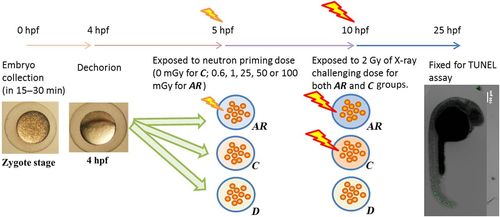
Schematic diagram showing the procedures for studying the radioadaptive response induced in zebrafish embryos that have been dechorionated at 4 hpf using a neutron priming dose and an X-ray challenging dose. AR: adaptive group, in which the dechorionated embryos received both the neutron priming dose and the X-ray challenging dose; C: adaptive control group, in which the dechorionated embryos were exposed to the X-ray challenging dose alone, without receiving a prior priming dose; D: dechorionated control group, in which the dechorionated embryos did not receive any radiation dose. |

Schematic diagram showing the procedures for studying the effect of X-ray photons on the radioadaptive response induced by alpha particles in zebrafish embryos that have been dechorionated at 4 hpf using a priming dose provided by (alpha particles) or (alpha particles + low-dose X-ray photons) and an X-ray challenging dose. AXY group: in which the dechorionated embryos received both the priming dose provided by (<0.88 mGy alpha-particle irradiation + level-Y X-ray irradiation) and the 2 Gy X-ray challenging dose, where level-Y was either 5 or 10 mGy; A group: in which the dechorionated embryos received both the priming dose provided by <0.88 mGy alpha-particle irradiation and the 2 Gy X-ray challenging dose; Control group: in which the dechorionated embryos were exposed to the X-ray challenging dose alone, without receiving a prior priming dose; D group: in which the dechorionated embryos did not receive any radiation dose. |

Representative images of stained embryos. (A) to (E): embryos from AR groups after first receiving a neutron priming dose of (A) 0.6 mGy, (B) 1 mGy, (C) 25 mGy, (D) 50 mGy and (E) 100 mGy, and then an X-ray challenging dose of 2 Gy; (F): a C group embryo after receiving an X-ray challenging dose of 2 Gy only; (G): a D group embryo without receiving any radiation dose. Images of embryos were captured by a confocal laser microscope with ×4 objective lens. A total of 15 to 25 sliced images with 25 µm intervals were captured for each embryo, which were then combined from top to bottom to generate the final image. |