
- Title
-
Development of a conditional liver tumor model by mifepristone-inducible Cre recombination to control oncogenic kras(V12) expression in transgenic zebrafish
- Authors
- Nguyen, A.T., Koh, V., Spitsbergen, J.M., Gong, Z.
- Source
- Full text @ Sci. Rep.
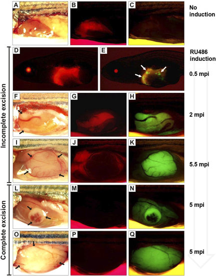
Mosaic pattern of Cre-mediated activation of EGFP-krasV12 in transgenic fish. Brightfield and corresponding fluorescence images of representative Triple-Tg fish are shown in the same rows. (A–C) Without induction, Triple-Tg fish showing normal liver morphology (A) with mCherry (B) but not EGFP-krasV12 (C). (D–K) Induced Triple-Tg fish at 1-month-old expressing both mCherry (D,G,J) and EGFP-krasV12 (E,H,K) in the liver, indicating the occurrence of incomplete Cre excision. After induction, many subsets of EGFP-positive liver cells were observed in 1.5-month-old fish (D,E). Liver tumors expressing EGFP developed in a 2.5-month-old (F,G,H) and 6-month-old Triple-Tg fish (I,J,K). (L–Q) Complete excision of the LChL cassettes observed in induced Triple-Tg fish at 6-month-old with the formation of large liver tumors (L,O) only expressing EGFP-krasV12 (N,Q) and no detectable mCherry fluorescence (M,P). Liver tumors are denoted by arrows. EXPRESSION / LABELING:
PHENOTYPE:
|

Heterogeneous liver tumors induced by krasV12. Histopathological examinations of liver tumors from Triple-Tg fish induced at 1-month-old. (A,B) Representative normal liver section from Triple-Tg fish without induction. (C,D) Liver tumor from induced Triple-Tg fish after 15 weeks displayed many vacuolated hepatocellular adenoma (HCA) with carcinoma grade 2 (HCC) arising in the center. (E,F) Liver tumor from induced Triple-Tg fish after 19 weeks showed HCC (grade 1) occupying 80% of the liver volume, with HCC (grade 2) arising centrally. (G,H) A Triple-Tg fish at 28 weeks after induction showing HCC (grade 2–3) involving the entire liver with extensive areas of hepatoblastoma (HB). Right panel showed high magnification of boxed area in the left panel. Arrows indicated the boundaries between different types of liver tumors. Scale bars, 100 µm. PHENOTYPE:
|
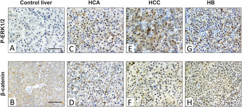
Deregulation of ERK and Wnt/β-catenin pathways in different types of krasV12-induced liver tumors. Three types of liver neoplasia including benign HCA, malignant HCC and HB were examined for the expression patterns of P-ERK1/2 and β-catenin via immunohistochemistry. (A,C,E,G) Strong mixed nuclear and cytoplasmic stainings of P-ERK1/2 were detected in three types of krasV12 liver tumors as compared to control liver of non-induced Triple-Tg fish. (B,D,F,H) Immunohistochemistry for β-catenin showed nuclear localization and accumulation of β-catenin only in HCC and HB, whereas normal liver and HCA displayed membranous staining of β-catenin. Scale bars, 50 µM. |