
- Title
-
Characterization of a novel zebrafish (Danio rerio) gene, wdr81, associated with cerebellar ataxia, mental retardation and dysequilibrium syndrome (CAMRQ)
- Authors
- Doldur-Balli, F., Ozel, M.N., Gulsuner, S., Tekinay, A.B., Ozcelik, T., Konu, O., Adams, M.M.
- Source
- Full text @ BMC Neurosci.
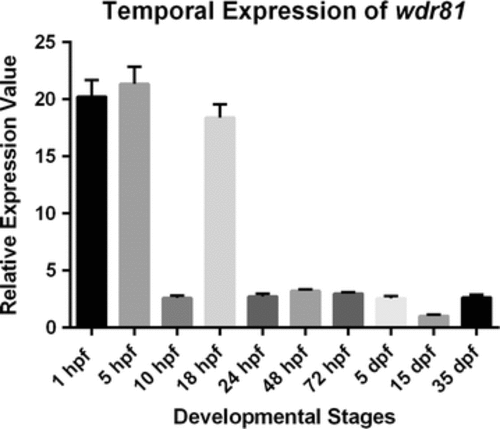
Relative temporal expression of wdr81 as determined by qRT-PCR. (1) 1 hpf embryo, (2) 5 hpf embryo, (3) 10 hpf embryo, (4) 18 hpf embryo, (5) 24 hpf embryo, (6) 48 hpf embryo, (7) 72 hpf larva, (8) 5 dpf larva, (9) 15 dpf larva, (10) 35 dpf juvenile zebrafish. Error bars represent +SE EXPRESSION / LABELING:
|
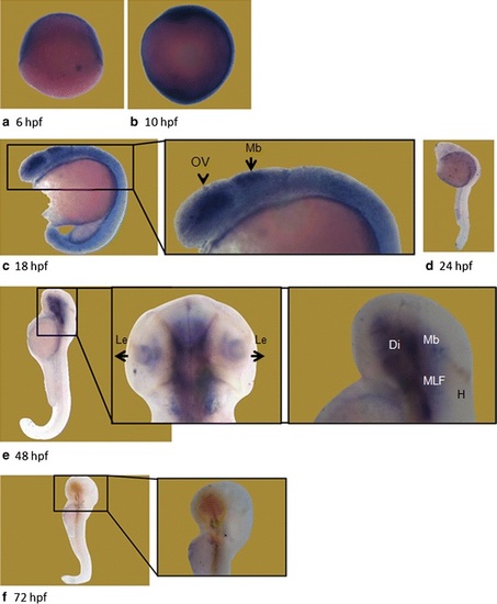
Whole mount in situ hybridization revealed differential expression of wdr81 transcript during embryonic development. Our results from the WMISH method are in parallel with the qRT-PCR data. The signal is high during the first 3 developmental timepoints (6–18 hpf), it is decreased and maintained during the rest of the development periods (24–72 hpf). OV optic vesicle, Mb midbrain, Le lens, H hindbrain, Di diencephalon, MLF medial longitudinal fascicle. EXPRESSION / LABELING:
|
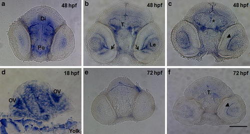
Transverse sections through the head regions of whole mount in situ hybridization specimens at 3 embryonic timepoints. The expression of wdr81 was observed in a regionally-specific manner by 48 hpf (a–c), whereas it was ubiquitously expressed at 18 hpf (d), and decreased by 72 hpf (e, f). Arrows indicate the optic nerve, asterisk the region of the nucleus of the medial longitudinal fascicle, and arrowhead the retina. Po preoptic area, Di diencephalon, T. midbrain tegmentum, Le lens, OV optic vesicle, Yolk yolk sac. Scale bar equals 100 μm EXPRESSION / LABELING:
|
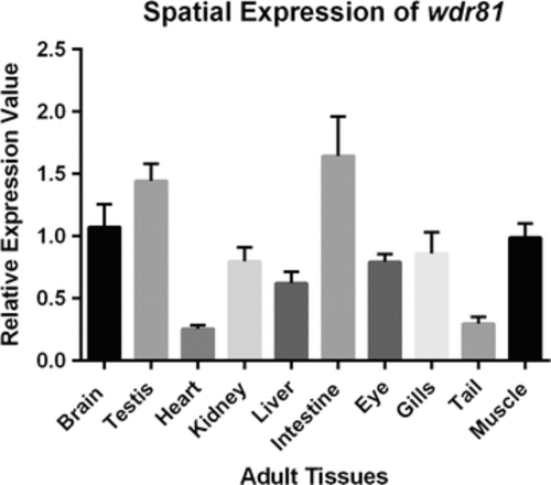
Relative spatial expression graphic of wdr81 as determined by qRT-PCR. (1) Brain, (2) testis, (3) heart, (4) kidney, (5) liver, (6) intestine, (7) eye, (8) gills, (9) tail, (10) muscle. Error bars indicate +SE |
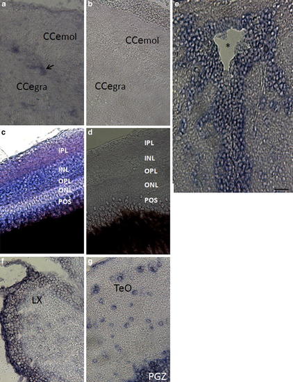
wdr81 expression in the adult brain and eye tissues. wdr81 expression was detected in the cerebellum (a), retina (c), tectal ventricle (e), brain stem (f), and optic tectum (g). Results with a sense probe in both cerebellum (b) and retina (d), which demonstrate no staining, indicates the specificity of the signal obtained with an antisense probe and both cerebellum (b) and retina (d) are shown. CCe mol Cerebellar molecular layer, Cce gra cerebellar granular layer, POS photoreceptor outer segments, ONL outer nuclear layer, OPL outer plexiform layer, INL inner nuclear layer, IPL inner plexiform layer, LX lobus vagus, TeO optic tectum, PGZ periventricular gray zone of the optic tectum. The arrow indicates the Purkinje cell layer and the asterisk the tectal ventricle. Scale bar equals 200 μm EXPRESSION / LABELING:
|