FIGURE SUMMARY
Title

"Slow" skeletal muscles across vertebrate species

Authors
Luna, V.M., Daikoku, E., Ono, F.
Source
Full text @ Cell Biosci.

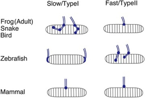
Schemas depicting motor neuron axons (blue) innervating individual muscle fibers. Note the differences in synaptic distribution between fiber types and animals

a Slow fibers (a single fiber shown in blue) are found directly underneath the skin. Synapses (shown in red) are found at the chevron-shaped body segment boundaries. At the individual fiber level, these synapses are at the distal edges. AChR pentamers found in these synapses do not include the λ or ε subunit, and generate currents with slow decay kinetics. b Fast fibers (a single fiber shown in blue) are located in layers deeper than slow fibers. Synapses (red) can be observed as round spots in each fiber. AChRs contain λ or ε subunit (yellow) and generate fast synaptic current kinetics

Acknowledgments
This image is the copyrighted work of the attributed author or publisher, and ZFIN has permission only to display this image to its users. Additional permissions should be obtained from the applicable author or publisher of the image. Full text @ Cell Biosci.