
- Title
-
Divergent evolution of two corticotropin-releasing hormone (CRH) genes in teleost fishes
- Authors
- Grone, B.P., Maruska, K.P.
- Source
- Full text @ Front. Neurosci.
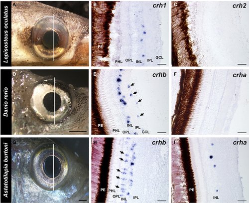
Localization of crh forms in the retina of fishes. Photographs of the eye of each species and adjacent 20 μm transverse sections of the retina reacted with each crh anti-sense probe are shown. (A–C) The spotted gar Lepisosteus oculatus shows crh1 expression in the inner nuclear layer, but crh2 is absent from the retina. (D–F) The zebrafish Danio rerio expresses crhb in the amacrine cell region of the inner nuclear and in the ganglion cell layer (arrows), but crha is absent from the retina. (G–I) The African cichlid fish Astatotilapia burtoni shows crhb label in two different cell types within the inner nuclear layer, amacrine and bipolar cells (arrows). crha was also found in the A. burtoni retina, but was restricted to the region of amacrine cells in the inner nuclear layer. GCL, ganglion cell layer; INL, inner nuclear layer; IPL, inner plexiform layer; OPL, outer plexiform layer; PHL, photoreceptor layer; PE, pigmented epithelium. Lines on (A,D,G) indicate the approximate position of the sections. Scale bars = 1 mm (A,D,G); 25 μm (B,C,E,F,H,I). EXPRESSION / LABELING:
|
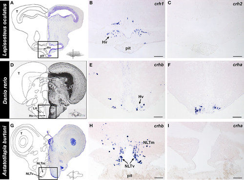
Localization of crh forms in the hypothalamus of fishes. (A) Representative transverse section of the spotted gar (Lepisosteus oculatus) brain stained with cresyl violet, with the approximate region of interest indicated (black box). (B) crh1-expressing cells were abundant in the ventral hypothalamus (Hv) of the gar, in the region above the pituitary (pit). (C) crh2 expression was absent from the hypothalamus of the spotted gar. (D) Representative transverse section from the zebrafish (Danio rerio) brain atlas (Wullimann et al., 1996), with the region of interest indicated (black box). (E) Zebrafish crhb-expressing cells were found in the nucleus lateralis tuberis (NLT) (labeled ventral hypothalamus, Hv, in atlas), but were also widely distributed throughout the brain. (F) Zebrafish crha expression was found exclusively in the NLT, and nowhere else in the brain. (G) Representative transverse section of the cichlid (Astatotilapia burtoni) brain stained with cresyl violet, with the approximate region of interest indicated (black box). (H) crhb-expressing cells were abundant in the NLT region above the pituitary gland of the cichlid. (I) crha expression was not detectable in the cichlid brain. Insets on (A,D,G) show lateral view of the brain with approximate location of transverse sections indicated (line). For each species, panels are adjacent sections labeled with different anti-sense crh probes. Hv, ventral zone of the periventricular hypothalamus; LH, lateral hypothalamic nucleus; NLTm, medial part of lateral tuberal nucleus; NLTv, ventral part of lateral tuberal nucleus; pit, pituitary; T, tectum. Scale bars = 100 μm (B,C); 25 μm (E,F,H,I). EXPRESSION / LABELING:
|