
- Title
-
Ewing Sarcoma Ewsa Protein Regulates Chondrogenesis of Meckel's Cartilage through Modulation of Sox9 in Zebrafish
- Authors
- Merkes, C., Turkalo, T.K., Wilder, N., Park, H., Wenger, L.W., Lewin, S.J., Azuma, M.
- Source
- Full text @ PLoS One

4 dpf MZ ewsa/ewsa mutants display an aberrant angle of Meckel’s cartilage and palatoquadrate. A. Lateral views (anterior to the left) of wt/wt (left) and MZ ewsa/ewsa (right) and ventral views of adult zebrafish. The calcified bones were visualized by alizarin red staining. B. Lateral views (anterior to the left) of (a) wt/wt and (b) MZ ewsa/ewsa and ventral views of (c) wt/wt and (d) MZ ewsa/ewsa chondrocytes from 4 dpf zebrafish embryos visualized with alcian blue. (e and f) Angle formed by Meckel’s cartilage showing that the palatoquadrate is wider in the MZ ewsa/ewsa mutant than wt/wt at 4 dpf and 7 dpf. bh: basihyal, d: dentary, m: Meckel’s cartilage, pq: palatoquadrate, ch: ceratohyal, cb: ceratobranchial. |
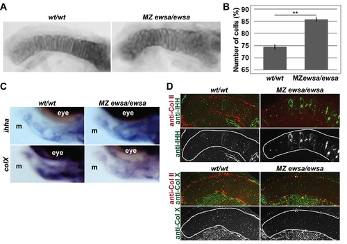
Prehypertrophic chondrocytes of Meckel’s cartilage in MZ ewsa/ewsa fails to differentiate into hypertrophic chondrocytes. A. Flat mounted Meckel’s cartilage of 4 dpf (left) wt/wt and (right) MZ ewsa/ewsa. B. Number of cells of Meckel’s cartilage of 4 dpf wt/wt and MZ ewsa/ewsa zebrafish (P = 0.0002). C. Lateral views (anterior to the left, dorsal to the top) of 4dpf (Left) wt/wt and (Right) MZ ewsa/ewsa visualized by in situ hybridization using probe for ihha (top panel) and colX (bottom panel). m: Meckel′s cartilage. D. Ventral views (anterior to the left) of Meckel′s cartilage of (Left) wt/wt and (Right) MZ ewsa/ewsa visualized by immunohistochemistry using (top) anti-IHH antibody (green), and anti-Collagen type II antibody (Red) (4dpf), and (bottom) anti-Collagen X antibody (green) and anti-Collagen type II antibody (Red) (5dpf). |

MZ ewsa/ewsa mutants display altered expression domains of Sox9 target genes. 27hpf of (Left) wt/wt and (Right) MZ ewsa/ewsa visualized by in situ hybridization using antisense RNA probe for A. sox5, B. noggin1, C. noggin2, D. bmp4, E. ctgfa, F. ctgfb, G. col2a1a and H. col2a1b. Top panel: low magnification images, middle and bottom panel: high magnification images. EXPRESSION / LABELING:
|
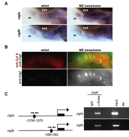
Ewsa binds to ctgfa and ctgfb target genes. A. Lateral views (anterior to the left, dorsal to the top) of 4dpf of (Left) wt/wt and (Right) MZ ewsa/ewsa visualized by in situ hybridization using probe for ctgfa (tom panel) and ctgfb (bottom panel). m: Meckel′s cartilage. B. Ventral views (anterior to the left) of 4dpf Meckel′s cartilage of (Left) wt/wt and (Right) MZ ewsa/ewsa visualized by immunohistochemistry using anti-Ctgf antibody (green), and anti-Collagen type II antibody (Red). C. (left) Schematics of ctgfa and ctgfb genes. Black circle: Sox9 binding site, black square: exon, arrows: PCR primer for ChIP assay. (right) ChIP assays were performed using 27 hpf zebrafish embryos. IgG: negative control of ChIP, anti-Ewsa: ChIP sample using EWSa antibody, input: 4.5% of DNA from total lysates was subjected to PCR, dw: negative control for PCR reaction. EXPRESSION / LABELING:
|
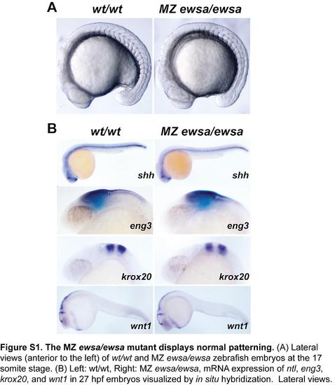
The MZ ewsa/ewsa mutant displays normal patterning. |

The MZ ewsa/ewsa mutant displays normal patterning of craniofacial chondrocytes. |
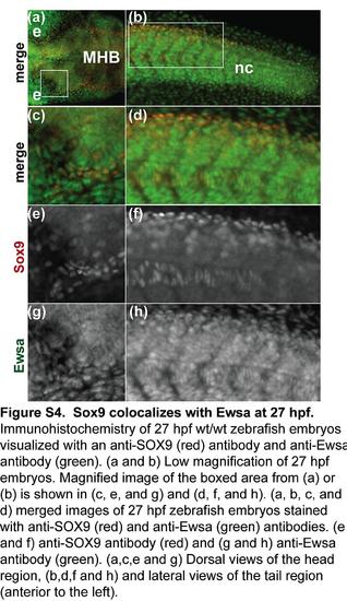
The Sox9 antibody recognizes zebrafish Sox9a. EXPRESSION / LABELING:
|