
- Title
-
Identification and expression analysis of zebrafish polypeptide α-N-acetylgalactosaminyltransferase Y-subfamily genes during embryonic development
- Authors
- Nakayama, Y., Nakamura, N., Kawai, T., Kaneda, E., Takahashi, Y., Miyake, A., Itoh, N., Kurosaka, A.
- Source
- Full text @ Gene Expr. Patterns

Temporal expression patterns of Y-subfamily galnt genes during zebrafish embryonic development. Zebrafish galnt cDNAs were amplified by RT-PCR at the indicated developmental stages. Cropped images of the gels are shown. The lowest panel shows results for zebrafish elongation factor 1-alpha (ef1a) as a control. These experiments were repeated three times and the data from a representative experiment are shown. EXPRESSION / LABELING:
|
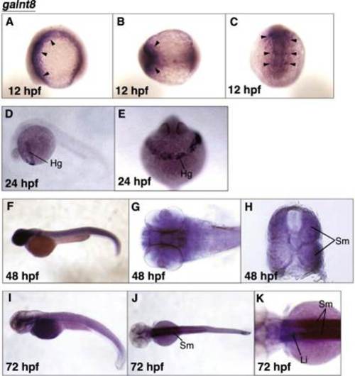
Spatial expression patterns of galnt8 during developmental stages. Whole mount in situ hybridization of galnt8 was performed at the developmental stages indicated at the bottom left corners. Lateral views with anterior to the left and dorsal to the top (A, D, F, and I). Dorsal views with anterior to the left (B, G, J, and K). A dorsal view of the head region (C). Anterior view of the hatching gland (E). A transverse section view of the trunk musculature (H). Arrowheads in A-C indicate the cephalic mesoderm. Hg; hatching gland, Sm; somitic musculature, Li; liver. EXPRESSION / LABELING:
|

Spatial expression patterns of galnt9 and galnt17 during developmental stages. Whole mount in situ hybridization of galnt9 (A–K) and galnt17 (L–X) was performed at the developmental stages indicated at the bottom left corners. Lateral views with anterior to the left and dorsal to the top (A, B, D, E, H, I, L, M, Q, R, U, and V). Dorsal views with anterior to the left (C, F, J, N, S, and W). Transverse section views of the middle hindbrain (G, K, O, T, and X) and of the trunk musculature (P). Te; telencephalon, Mb; midbrain, Hb; hindbrain, Re; retina, Di; diencephalon, Ov; otic vesicle, Sc; spinal cord, Sm; somitic musculature. EXPRESSION / LABELING:
|

Spatial expression patterns of galnt18a and galnt18b during developmental stages. Whole mount in situ hybridization of galnt18a (A–N) and galnt18b (O–Z) was performed at the developmental stages indicated at the bottom left corners. Lateral views with anterior to the left and dorsal to the top (A, C, D, E, F, G, J, K, O, P, S, and W). Dorsal views with anterior to the left (B, H, L, Q, T, and Y). Transverse section views of the trunk musculature (I), the middle hindbrain (M, V, and Z), and the posterior hindbrain (N). Te; telencephalon, Di; diencephalon, Hb; hindbrain, Sm; somitic musculature, Mb; midbrain, Ol; olfactory vesicle, Ov; otic vesicle, Pg; pineal gland. Arrowheads and arrows indicate galnt18a expression at the cleavage furrow (A and B), and in the dorsal posterior medulla oblongata (K and N), respectively. EXPRESSION / LABELING:
|

Maternal expression of galnt18a. Zebrafish galnt cDNAs were amplified by RT-PCR at the 1-2-cell stage and 24 hpf. Cropped images of the gels are shown. The lowest panel shows results for zebrafish elongation factor 1-alpha (ef1a) as a control. These experiments were repeated three times and the data from a representative experiment are shown. |
Reprinted from Gene expression patterns : GEP, 16(1), Nakayama, Y., Nakamura, N., Kawai, T., Kaneda, E., Takahashi, Y., Miyake, A., Itoh, N., Kurosaka, A., Identification and expression analysis of zebrafish polypeptide α-N-acetylgalactosaminyltransferase Y-subfamily genes during embryonic development, 1-7, Copyright (2014) with permission from Elsevier. Full text @ Gene Expr. Patterns