
- Title
-
An assessment of the long-term effects of simulated microgravity on cranial neural crest cells in zebrafish embryos with a focus on the adult skeleton
- Authors
- Edsall, S.C., Franz-Odendaal, T.A.
- Source
- Full text @ PLoS One

Melanophores on the dorsal head of a larval zebrafish. A) before treatement with epinephrine. B) post treatment. The same fish is shown in A and B. B) shows the region in which melanophores were counted. Scale bars are 200 μm. |

Landmarks for morphometric analyses. A) The five landmarks assigned to the dissected right opercle of an adult fish. B) Ventral view of the cartilage-stained pharyngeal arches of a SMG specimen showing the 46 landmarks used, SL 4.0 mm. Scale bars are 500 μm and 100 μm in A and B respectively. |

Adult skeletal phenotypes of fish exposed to SMG as embryos. Fish are bone stained with Alizarin red. A–H) Left lateral views, (I–P) dorsal views. All fish are four months old. Scale bars are 500 μm. |
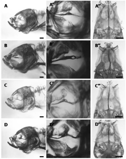
The parasphenoid phenotypes in adult bone-stained zebrafish exposed to 24 hours of SMG starting at 12 hpf. A–D) left lateral views. Higher magnifications are given in A′-D′ and in ventral view in A′′-D′′. A) slight thickening of the parasphenoid in the region where it articulates with the orbitosphenoid, 14.0 mm SL. B) thickened parasphenoid with a hole in the posterior region, 15.0 mm SL. C) parasphenoid is slightly thickened in the region where it articulates with the orbitosphenoid and severely bent posteriorly, 15.0 mm SL. D) parasphenoid is thickened at the orbitosphenoid articulation and bent towards the posterior end, 14.0 mm SL. All scale bars are 500 μm. |

Left lateral view of acid-free double-stained juveniles from the 24 h SMG at 12 hpf group. A) no buckling in the parasphenoid at 10 dpf, 3.3 mm SL. B) no buckling of the parasphenoid at 35 dpf, 7.5 mm SL. C) severe buckling in the parasphenoid at 65 dpf, 14 mm SL. Scale bars are 500 μm. |

Morphometric analyses of the opercle. A) Right opercle from a 24 h SMG at 12 hpf adult fish showing buckling in the anterior-posterior direction (arrows); dashed line indicates the normal shape of this bone. B) vector analysis comparing the control consensus (CNV, arrow origins) to the consensus for this SMG group (arrow end points). |