
- Title
-
Optimization and in vivo toxicity evaluation of G4.5 PAMAM dendrimer-risperidone complexes
- Authors
- Prieto, M.J., del Rio Zabala, N.E., Marotta, C.H., Carreño Gutierrez, H., Arévalo Arévalo, R., Chiaramoni, N.S., del Valle Alonso, S.
- Source
- Full text @ PLoS One
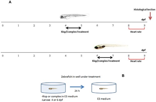
Timeline representing the stage specificity of the effects of risperidone and DG4.5-Risp in developing zebrafish. Larvae were exposed to 5 µM risperidone or DG4.5-Risp for 24-h periods from 4 dpf or 6 dpf and subsequently rescued into a conditioned E3 medium (A). Schematic representation of the in vivo treatment (B). |
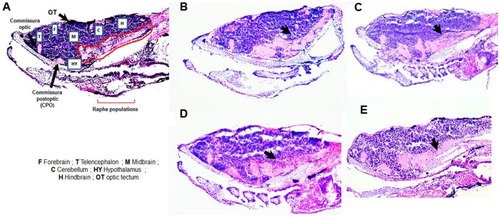
Images of histological sections of brain tissue stained with hematoxylin-eosin. A) control, B) risperidone (Risp) at 4 dpf, C) Risp at 6 dpf, D) DG4.5-Risp at 4 dpf, and E) DG4.5-Risp at 6 dpf. Larvae were analyzed three times (n = 3) at 10 dpf. |
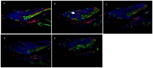
Immunohistochemistry images of brain tissue. Tyrosine hydroxylase, labeled with Cy2 (green), and Calretinin, labeled with Cy3 (red). A) control, B) risperidone (Risp) at 4 dpf, C) Risp at 6 dpf, D) DG4.5-Risp at 4 dpf, and E) DG4.5-Risp at 6 dpf. Larvae were analyzed three times (n = 3) at 10 dpf. |
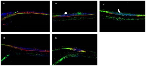
Immunohistochemistry images of spinal cord tissue. Tyrosine hydroxylase, labeled with Cy2 (green), and Calretinin, labeled with Cy3 (red). A) control, B) risperidone (Risp) at 4 dpf, C) Risp at 6 dpf, D) DG4.5-Risp at 4 dpf, and E) DG4.5-Risp at 6 dpf. Larvae were analyzed three times (n = 3) at 10 dpf. |