
- Title
-
The connexin 30.3 of zebrafish homologue of human connexin 26 may play similar role in the inner ear
- Authors
- Chang-Chien, J., Yen, Y.C., Chien, K.H., Li, S.Y., Hsu, T.C., Yang, J.J.
- Source
- Full text @ Hear. Res.
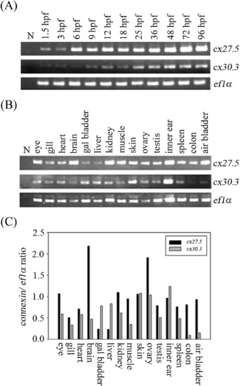
Zebrafish cx27.5 and cx30.3 transcripts expressed during embryo development stage and in adult tissues. (A) The expression of the zebrafish cx27.5 and cx30.3 gene during embryonic stage. Total RNAs from various stages of zebrafish were used as template for RT-PCR. The cx27.5 gene expressed at low levels in the 1.5 to 3 hpf and dominantly expressed after 6-96 hpf. In contrast, cx30.3 transcripts were hardly detected until 9 hpf. (B) Total RNAs from various organs of zebrafish were conducted to use as template for RT-PCR. Each bar shows the expression of cx27.5 or cx30.3 transcripts in various tissues of adult zebrafish relative to the expression of zebrafish elongation factor 1-alpha (ef1α). The ef1α was used as an internal control. (C) We found that the cx27.5 gene expressed in most adult tissues, except liver and gall bladder tissues. Meanwhile, the cx30.3 gene dominantly expressed in the cells of ovary, skin and inner ear of zebrafish. hpf: hour post fertilization, N: negative control. EXPRESSION / LABELING:
|
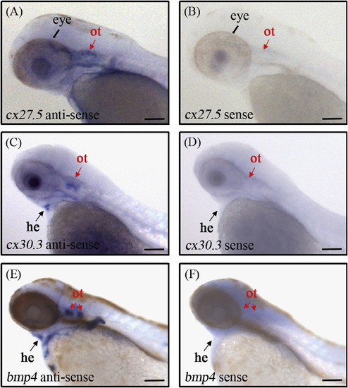
WISH of zebrafish cx27.5 and cx30.3 mRNA in 72 hpf embryos. Embryos at 72 hpf were stained with antisense riboprobe by WISH. All images were lateral views, anterior is to the left and dorsal is up. (A, B) The zebrafish cx27.5-specific signal was weakly detected in otic vesicle (ot; red arrows) and appeared to be more superficial. In addition, cx27.5 signal was also detected in the eye (broad black arrows). (C, D) The cx30.3-specific signal was detected in otic vesicle and heart (he; black arrows) of 72 hpf embryos. (E, F) bmp4 mRNA probe was used as an otic vesicle marker. Scale bars represent 0.1 mm. (For interpretation of the references to color in this figure legend, the reader is referred to the web version of this article.) EXPRESSION / LABELING:
|

Zebrafish Cx30.3 proteins expressed in the heart and inner ear of 72 hpf embryos. The sections from paraffin-embedded 72 hpf embryos were immunodetected with antibodies against Cx30.3 and counterstained using HE staining. Zebrafish Cx30.3 proteins, indicated by yellow arrows, were shown in the macular cells of inner ear (A-D) and in the heart (E-H) using anti-Cx30.3 antibody. However, Cx30.3 proteins were not detected by pre-immune serum (I-K). (A, E, I) Sections from the 72 hpf embryos were counterstained with DAPI to highlight the nuclei. (B, F, J) Cx30.3 proteins expression. (C, G, K) Panel A, E or I merges with panel B, F or J respectively. (D, H) HE staining. ot: otic vesicle. Scale bars represent 50 µm. (For interpretation of the references to color in this figure legend, the reader is referred to the web version of this article.) EXPRESSION / LABELING:
|
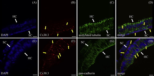
Zebrafish Cx30.3 proteins expressed in the cell membrane of hair cells of adult inner ear. Zebrafish Cx30.3 protein is expressed in the cell membrane of hair cells of the adult inner ear. Confocal images of sections from the adult inner ear that were immunostained with antibodies against zebrafish Cx30.3, acetylated tubulin (a hair cell marker) and pan-cadherin (a cell membrane marker) are displayed. The Cx30.3 protein, indicated by yellow arrows, is expressed at higher levels in supporting cells (SC) than in hair cells (HC) of maculae (A-D). In addition, the Cx30.3 protein localized to the cell membrane of hair cells (E-H). Sections from the adult inner ear were counterstained with DAPI to highlight the nuclei. Scale bars represent 10 µm. (For interpretation of the references to color in this figure legend, the reader is referred to the web version of this article.) EXPRESSION / LABELING:
|
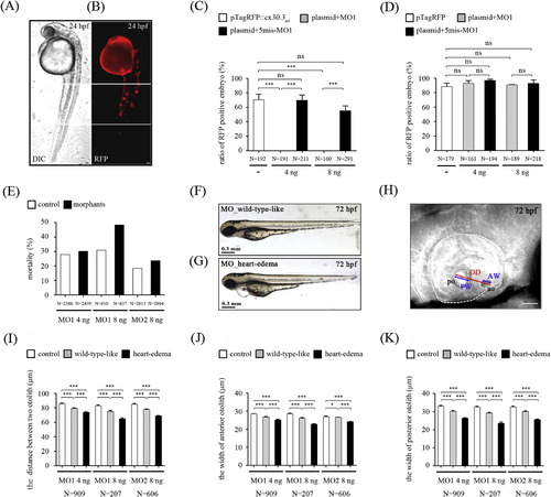
Morpholino knockdown of zebrafish cx30.3 expression in zebrafish embryos. (A) Either TagRFP proteins or TagRFP-tagged Cx30.3 fusion proteins was injected at the one-cell stage embryo in the presence or absence of morpholinos. Injected-embryos were observed at 24 hpf using fluorescence microscopy. (B) The mosaic expression pattern of TagRFP (fusion) proteins was shown in plasmid-injected embryos. (C) The ratio of Cx30.3-TagRFP positive embryos in the presence or absence of ATG-MO1 (MO1). The translation-blocking MO1 was able to completely knockdown the expression of Cx30.3 fusion proteins in both 4 ng and 8 ng morpholinos-injected embryos, whereas the negative control morpholino with 5 nucleotides substitution (5mis-MO1) had no significant effect. (D) Neither MO1 nor 5mis-MO1 was able to significantly reduce the TagRFP expression in embryos. Therefore, MO1 and splicing-inhibiting morpholino e1i1-MO2 (MO2) were injected respectively to in vivo knockdown cx30.3 transcripts in zebrafish embryos. The cx30.3 morphants, injected with 4 ng or 8 ng morpholinos (MO1 and MO2), showed the higher mortality (E) and mild alternation in their phenotype, termed wild-type-like (F) and heart edema (G), compared with the control-injected embryos at 72 hpf. All DIC images are lateral views, anterior is to the left and dorsal is up. Scale bars represent 0.3 mm. (H) The distance between two otoliths (OD) and the width of each otolith (anterior otolith, AW; posterior otolith, PW) were then measured at 72 hpf. The schematic presentation of the measurement in otic vesicle (white dotted line) was shown. Scale bars represent 50 µm. The statistical analyses visualize the significant reduction in the distance between two otoliths (n = 303 in each group) (I) as well as the width of anterior otolith (n = 69 in each group) (J) or posterior otolith (n = 202 in each group) (K) in both morphants (wild-type-like and heart edema). All data were presented as the mean ± SD and were considered significant difference for P < 0.05, P < 0.01 and P < 0.001 as compared with the control group. Overall, zebrafish cx30.3 morphants showed the wild-type-like and heart-edema phenotypes with smaller inner ear. ‘N’ represents the number of injected embryos. ao: anterior otolith, po: posterior otolith, AW: the width of anterior otolith, PW: the width of posterior otolith, OD: the distance between two otoliths. PHENOTYPE:
|
Reprinted from Hearing Research, 313, Chang-Chien, J., Yen, Y.C., Chien, K.H., Li, S.Y., Hsu, T.C., Yang, J.J., The connexin 30.3 of zebrafish homologue of human connexin 26 may play similar role in the inner ear, 55-66, Copyright (2014) with permission from Elsevier. Full text @ Hear. Res.