
- Title
-
TAILOR: transgene activation and inactivation using lox and rox in zebrafish
- Authors
- Park, J.T., and Leach, S.D.
- Source
- Full text @ PLoS One

Heterospecific recombination of rox and lox sites by Dre and Cre in zebrafish embryos. (A, B) Schematic of ubb-Dre (A) and ubb-Cre (B) driver lines and corresponding Rox-Nuc-mCherrys-Rox and Lox-Nuc-mCherry-Lox reporters. Additional cryaa:Venus cassette facilitates identification of transgene-expressing embryos. Open triangles indicate Tol2 arms. (C) Images from double transgenic embryos produced by indicated crosses of ubb-Dre and ubb-Cre driver lines with either Rox-Nuc-mCherry-Rox-eGFP or Lox-Nuc-mCherry-Lox-eGFP reporter lines. Activation of eGFP confirms Dre-specific recombination of Rox-Nuc-mCherry-Rox reporter and Cre-specific recombination of Lox-Nuc-mCherry-Lox reporter. Scale bar: 200 μm. |

Induction of DrePR recombinase activity by RU486. (A) Schematic of ubb-DrePR driver line and Rox-Nuc-mCherry-Rox reporter. Additional cryaa:eCFP cassette facilitates identification of transgene-expressing embryos. Open triangles indicate Tol2 arms. (B) Tight control of DrePR recombinase activity by RU486. ubb-DrePR; Rox-Nuc-mCherry-Rox-eGFP embryos were treated with and without 4 μM RU486 between 24 and 48 hpf, and imaged at 96 hpf. No expression of eGFP is observed in untreated (-RU486) or tamoxifen (4-OHT)-treated embryos, while treatment with RU486 results in potent induction of eGFP expression indicating successful recombination of Rox-Nuc-mCherry-Rox-eGFP allele. (C) To quantify the efficiency of Dre recombination in Dre-expressing embryos and DrePR recombination in DrePR-expressing embryos, the intestine of larval zebrafish (4 dpf) were dissected following treatment with RU486 at the indicated concentration between 24–48 hpf. DAPI-labeled cells also labeled by either nucleus mCherry or cytoplasmic eGFP were counted. Maximal recombination frequency is achieved an an RU486 concentration of 4 μM, at a level comparable with Dre lacking the PR fusion. Scale bar: 25 μm. |

Combinatorial activation of DrePR and CreERT2. (A) Schematic of ubb:lox-stop-lox-rox-Nuc-mCherry-stop-ro x-eGFP dual reporter for assessment of both Cre- and Dre-mediated recombination, along with ubb-CreERT2 and ubb-DrePR driver lines. Ocular and cardiac fluorescence conveyed by additional cryaa:mCherry, cmlc2:eGFP and cryaa:eCFP cassettes facilitates identification of transgene-expressing embryos. Open triangles indicate Tol2 arms. (B) Triple transgenic fish were treated for 24 hrs with or without 4-OHT and RU486 as indicated, and imaged at 96 hpf. While untreated embryos showed no transgene-specific fluorescence besides that provided by the ocular mCherry, ocular eCFP and cardiac eGFP markers (Fig. 3B a1, a2, a3, and a4), embryos treated with only 4-OHT displayed widespread activation of nuc-mCherry, but no activation of eGFP (Fig. 3B b1, b2, b3, b4). In contrast, embryos simultaneously treated with both 4-OHT and RU486 displayed expression of both nuc-mCherry and eGFP (Fig. 3B c1, c2, c3, and c4). Scale bar: 200 µm. (C) Confocal imaging of dissected intestine, liver, and pancreas, confirming patterns of mCherry and eGFP expression observed in whole embryos. Following combined treatment with 4-OHT and RU486, a majority of cells in each tissue express either nuclear mCherry or cytoplasmic eGFP, with a smaller fraction of cells expressing both. Scale bar: 25 μm. |
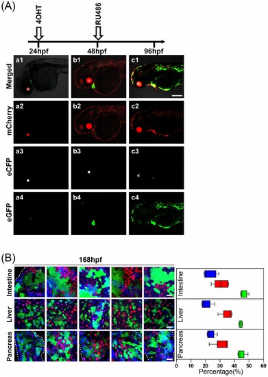
Sequential transgene activation and inactivation using lox and rox (TAILOR). (A) ubb:lox-stop-lox-rox-Nuc-mCherry-stop-ro x-eGFP;ubb-CreERT2; ubb-Dre triple transgenic fish were subjected to treatment with 4-OHT beginning at 24 hpf, followed by removal and replacement with RU486 at 48 hpf. The untreated triple transgenic embryos (24 hpf) showed no transgene-specific fluorescence besides that provided by the ocular mCherry, ocular eCFP and cardiac eGFP markers (Fig. 4A a1, a2, a3, and a4). The effective induction of nuclear mCherry expression was observed following 24 hrs of 4-OHT treatment (Fig. 4A b1 and b2). Following staged treatment with RU486 initiated at 48 hpf and left in place for 24 hrs, effective activation of eGFP expression was observed (Fig. 4A c1 and c4). Scale bar: 200 μm. (B) Quantification of relative numbers of intestinal, liver and pancreatic cells expressing mCherry (red), eGFP (green) or neither (blue) at 7 days following sequential 4OHT and RU486 exposure as above. Note high fraction of cells undergoing sequential CreERT2-mediated activation and DrePR-mediated inactivation of mCherry expression, as indicated by eGFP expression. Scale bar: 25 μm. |