
- Title
-
A Phenotypic Screen in Zebrafish Identifies a Novel Small-Molecule Inducer of Ectopic Tail Formation Suggestive of Alterations in Non-Canonical Wnt/PCP Signaling
- Authors
- Gebruers, E., Cordero-Maldonado, M.L., Gray, A.I., Clements, C., Harvey, A.L., Edrada-Ebel, R., de Witte, P.A., Crawford, A.D., and Esguerra, C.V.
- Source
- Full text @ PLoS One
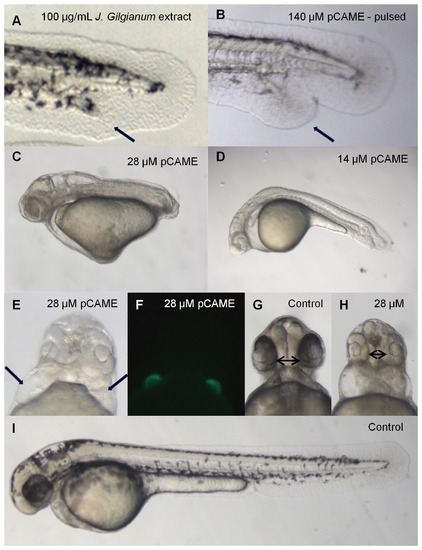
Phenotypic characterization of pCAME-treated embryos. (A) Embryo treated with 100 μg/mL crude methanolic extract of J. gilgianum. Black arrow denotes ectopic tail. Unless otherwise noted, all treatments were performed at 2- to 4-cell stages. (B) Embryo treated with 140 μM pCAME pulsed 1h at tailbud stage, black arrow denotes ectopic tail; (C) pCAME-treated embryo with 28 μM and with (D) 14 μM pCAME; (E) Embryo treated with 28 μM pCAME; black arrows denote 2 hearts; (F) cmlc2:eGFP embryo treated with 28 μM pCAME; (H) Embryo treated with 28 μM pCAME; black double arrow indicates synophthalmia; (G) and (I) Vehicle-treated control (1% DMSO), black double arrow denotes normal distance between eyes. All embryos are at 48 hpf. The main phenotypic characteristics of pCAME-treated embryos are: tail duplication, AP-axis shortening, absence of pectoral fins, decrease in pigmentation, body curvature, synophthalmia and cardia bifida. |
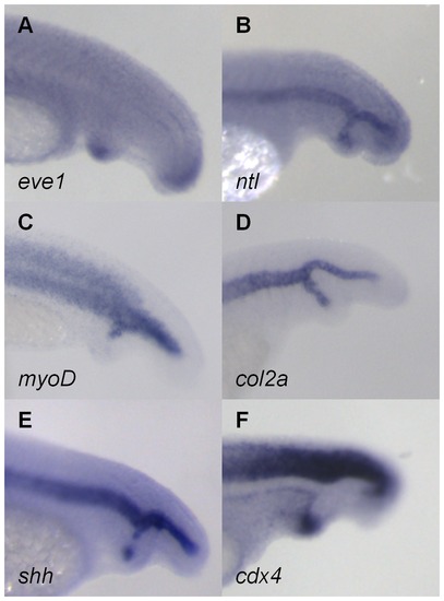
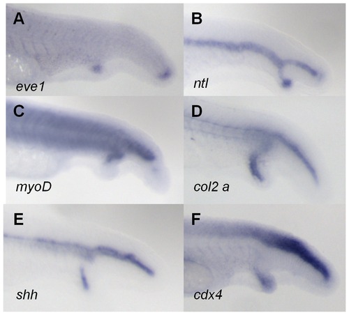
Analysis of tissue-specific marker expression in pCAME-induced ectopic tails. (A) eve1; (B) ntl; (C) myoD; (D) col2a; (E) shh; and (F) cdx4 expression. All embryos were pulsed with 140 μM pCAME for 1h at tailbud stage and fixed at 30 hpf. Lateral views. |
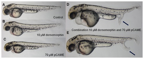
Synergy between pCAME and dorsomorphin. (A) Wild type pulsed with 1% DMSO in Danieau’s solution; (B) 10 µM dorsomorphin pulse; (C) 70 μM pCAME pulse; (D) and (E) Combination of 10 μM dorsomorphin and 70 μM pCAME pulse. Black arrows denote ectopic tails. All AB embryos are at 48 hpf and pulsed for 1h at tailbud stage. A significant increase of the percentage of ectopic tail formation was observed as well as a more pronounced phenotype. |
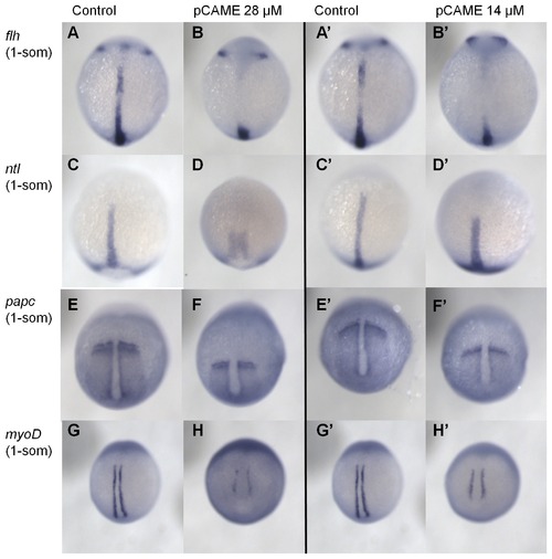
pCAME induces convergence and extension defects. (A) and (B) flh expression; (C) and (D) ntl; (E) and (F) papc; (G) and (H) myoD. All embryos were treated at 2-4 cell stages with 14 or 28 μM pCAME and fixed at 1 somite stage. Dorsal view. Expression domains of compound-treated fish are altered as would be predicted for CE defects. Embryos have a broader and shorter notochord and impaired convergence and extension of the para-axial mesoderm and the somites. |
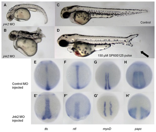
jnk2 morpholino knockdown induces convergence extension defects. (A) and (B) 2 mM jnk2 MO-injected fish A: 38 % B: 9 %; (C) Wild type; (D) 150 μM SP600125 pulsed for 1 h at tailbud stage. All embryos are at 48 hpf. Black arrow denotes duplicated tail. (E-H) Control MO injected fish; (E′-H′) jnk2 MO injected fish; (E) flh; (F) ntl; (G) myoD; (H) papc. |
Tissue marker analysis in SP600125-induced ectopic tails. (A) eve1; (B) ntl; (C) myoD; (D) col2a; (E) shh; and (F) cdx4 expression. All embryos were pulsed with 140 μM pCAME for 1h at tailbud stage and fixed at 30 hpf. Lateral views. |