FIGURE SUMMARY
Title

Zebrafish models for ectopic mineralization disorders: practical issues from morpholino design to post-injection observations

Authors
Hosen, M.J., Vanakker, O.M., Willaert, A., Huysseune, A., Coucke, P., and De Paepe, A.
Source
Full text @ Front. Genet.

Binding of the anti-sense morpholino chain to the RNA chain. MOs have standard nucleic acid bases bound to morpholino ring, which are linked through phosphorodiamidate groups, while RNA has ribose rings which are linked with phosphates.

Developmental stages of zebrafish, from zygote to adult. Zygote: the newly formed fertilized egg after completion of the first zygotic cell cycle. Cleavage: zygotic cell cycles 2–7 occur rapidly and synchronously. Blastula: rapid and metasynchronous cell cycles (8, 9) occur, which give way to lengthened, asynchronous ones at the midblastula transition, then epiboly begins. Epiboly is the first coordinated cell movement in zebrafish embryos and begins before gastrulation. Gastrula: morphogenetic movements of involution, convergence, and extension from the epiblast, hypoblast, and embryonic axis through the end of epiboly occur. Bud-100% epiboly is the stage where epiboly completely covers the yolk plug. Segmentation: Somites (after completion of epiboly and initial appearance of the tail bud, first the somatic furrow forms and makes a boundary, between what will become the first and second somites), pharyngeal arch primordia, and neuromeres develop, primary organogenesis and earliest movements take place, and the tail appears. Pharyngula: phylotypic stage of embryo, body axis straightens from its early curvature around the yolk sac; circulation, pigmentation, and fins begin development. Hatching: completion of rapid morphogenesis of primary organ systems, cartilage development in head and pectoral fin, hatching occurs asynchronously across individuals. Larval: swim bladder inflates; food-seeking and active avoidance behaviors.

Evaluation of the effect of SJ MO in the PCR product. Hypothetical representation of intron retention and exon skipping as a consequence of SJ MO injection. (A) MO is placed on exon2/intron2 border on targated genes pre-mRNA. The result can show (B) retention of intron 2, or (C) skipping of exon 2. FW, forward primer; REV, reverse primer.

Calcein staining of 6 dpf embryos showing staining of the ceratobranchials 5 (cb5), cleithrum (cl), dentary (d), entopterygoid (en), opercular bone (op), and parasphenoid (ps), in the skull, and anterior tip of the notochord (no).

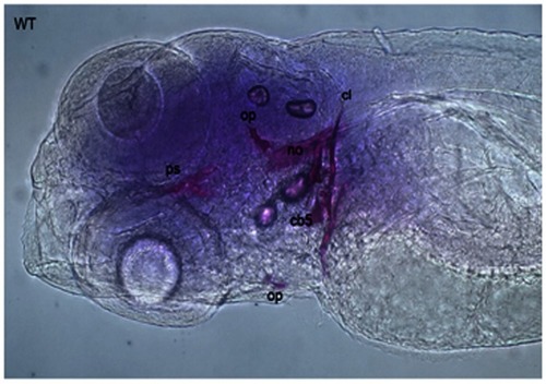
Alizarin Red staining of a 5 dpf embryo showing cleithrum (cl), opercular bone (op), parasphenoid (ps), and ceratobranchials 5 (cb) with a set of three teeth.

MicroCT scanning of whole 4 dpf embryos. The white spot in the head region represents a signal for the mineralized otoliths.

Transferase dUTP nick end labeling staining of 5 dpf embryos. More fluorescent dots are observed in the tail region of Abcc6a-MO injected fish (right) demonstrating more apoptosis compared to the tail region of the wild-type fish (left).

Detection of mitochondrial membrane potentiality on 2 dpf embryos by MitoTracker Red CM-H2XRos staining. (A) Control fish showing no fluorescent staining, and (B) staining with 500 nM MitoTracker Red CM-H2XRos for 2 h showing fluorescent staining of mitochondria in the head region of a mitochondrial disease model.

Overview of MO injection and post-injection follow-up to study ectopic mineralization zebrafish models.

Acknowledgments
This image is the copyrighted work of the attributed author or publisher, and ZFIN has permission only to display this image to its users. Additional permissions should be obtained from the applicable author or publisher of the image. Full text @ Front. Genet.