
- Title
-
Quantitative Genetic Analysis of Retinal Degeneration in the Blind Cavefish Astyanax mexicanus
- Authors
- O'Quin, K.E., Yoshizawa, M., Doshi, P., and Jeffery, W.R.
- Source
- Full text @ PLoS One
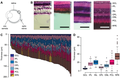
Variation in retinal thickness among Astyanax F2 hybrids. (A) Diagram of the eye, retina, and retinal landmarks. Black box indicates an example region where we recorded retinal thickness. (B) Example retinal sections from the eyes of four F2 hybrids. Scale bar denotes 50 µm and brackets denote individual retinal layers. Cavefish-like retinas (first two panels) were generally thin and may be missing some layers entirely, including the photoreceptor cell layer. Surface fish-like retinas (last two panels) were generally thicker with layers that are well-differentiated and clearly laminated. In some individuals, the retinal pigment epithelium (RPE) was hypopigmented (first and third panels). (C) Retinal thickness among 115 Astyanax SFxCF F2 hybrids. Yellow bars denote RPEs that were hypopigmented. (D) Box-plots illustrate variance in the thickness of individual retinal layers. The retinal layers shown are: ganglion cell layer (GCL), inner plexiform layer (IPL), inner nuclear layer (INL), outer plexiform layer (OPL), outer nuclear layer (ONL), photoreceptor cell layer (PCL), and retinal pigment epithelium (RPE). |