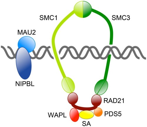
Overview of the cohesin complex and its associated proteins. The cohesin complex consists of four core subunits: SMC1, SMC3, RAD21, and SA. Together these subunits form a large ring capable of topologically encircling DNA strands. Other proteins regulate cohesin’s binding to DNA and its residency there. The NIPBL/MAU2 dimer loads cohesin onto DNA, whereas WAPL/PDS5 release cohesin from chromosomes by opening the SMC3-RAD21 interface.
Comparison of the top 200 affected probe sets in zebrafish embryos depleted of different cohesin subunits. Venn diagrams showing the overlap of the top 200 probe sets affected in zebrafish esco2 and nipbl morphants, and smc1ahi1113a and rad21nz171 mutants at 24 and 48 hpf (q value < 0.05).
Flat-mount staining (anti-HNK-1) of trigeminal ganglia in wild type (left) and rad21 mutant (right) zebrafish embryos. In rad21 mutants, central neuronal clumping occurred (arrow), and axons failed to migrate and populate anterior regions (red oval).
Model for diverse cohesinopathy phenotypes. In interphase (red shading), cohesin binding to chromatin is dynamic, with varying residency times. Interphase cohesin binding is likely to be cell type-specific and to contribute toward regulating developmental genes. Mutations in cohesin subunits and their key interphase regulators (e.g., Nipbl, Hdac8) primarily impact on the regulation of gene expression, including transcriptional regulation of growth pathways. This results in syndromic developmental defects that derive from dysregulated transcription, with the possibility of cell death as a contributing factor. From S phase to G2/M (blue shading), the overriding function of cohesin involves sister chromatid cohesion and DNA damage repair. Key regulators in this process include the CoAT ESCO2 and other DNA damage repair proteins. Mutations in these regulators result in chromosome segregation defects, genomic instability, and cell death. Increased cell death and reduced cell proliferation results in too few cells to make up body structures, leading to a different class of developmental defects and dysregulation of metabolic pathways. Transcription of a small subset of hypersensitive genes, including some in the Notch signaling pathway, appears to be sensitive to both interphase and S/G2 modes of cohesin binding.
Acknowledgments
This image is the copyrighted work of the attributed author or publisher, and
ZFIN has permission only to display this image to its users.
Additional permissions should be obtained from the applicable author or publisher of the image.
Full text @ Front. Genet.
Your Input Welcome
Thank you for submitting comments. Your input has been emailed to ZFIN curators who may contact you if
additional information is required.
Oops. Something went wrong. Please try again later.