
- Title
-
In Vivo Chemical Screening for Modulators of Hematopoiesis and Hematological Diseases
- Authors
- Zhang, Y., and Yeh, J.R.
- Source
- Full text @ Adv. Hematol.
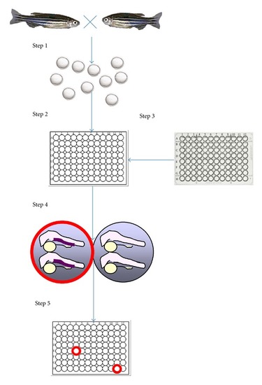
Chemical screening using zebrafish embryos. Step 1—Wild-type, reporter, or mutant zebrafish are crossed to obtain embryos. Step 2—Once reaching an investigator-specified developmental stage (usually between 0–5 days after fertilization), embryos are arrayed into multi-well plates either manually or by automation. Step 3—Compounds from a chemical library are added into the wells containing the embryos using a multichannel pipette or a pin-transfer device. Step 4—After reaching the developmental stage for phenotype manifestation, which is usually within hours to a couple of days after the compound treatment, embryos may be subjected to staining procedures, reporter, or functional assays to detect chemical-induced phenotypes or reversal of genetic phenotypes. The images shown here depict differential hematopoietic gene expression between the compound-treated (red circle) and vehicle-treated (black circle) embryos as detected by RNA |