
- Title
-
Zebrafish chemical screening reveals the impairment of dopaminergic neuronal survival by cardiac glycosides
- Authors
- Sun, Y., Dong, Z., Khodabakhsh, H., Chatterjee, S., and Guo, S.
- Source
- Full text @ PLoS One
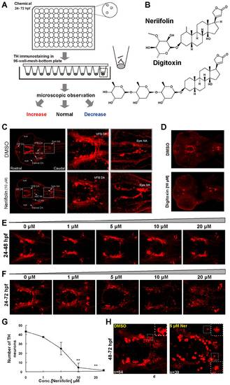
Zebrafish chemical screen identifies Neriifolin, a member of cardiac glycoside family, which disrupts the pattern of DA neurons in the ventral forebrain. (A) Schematic diagram of the chemical screening platform, through which Neriifolin was identified as a hit that decreases ventral forebrain DA neurons. (B) Structure of two cardiac glycosides, Neriifolin and Digitoxin, both of which disrupt the pattern of VFB DA neurons. (C) Embryos treated with 10 μM Neriifolin showed a decrease of VFB DA neurons (middle panels), whereas the Sym NA neurons were normal (right panels). (D) Treatment with another cardiac glycoside, Digitoxin, similarly decreased VFB DA neurons but not Sym NA neurons. (E) Embryos treated with different concentrations of Neriifolin from 24 hpf to 48 hpf showed no obvious defect in the pattern of VFB DA neurons. (F) Embryos treated with different concentrations of Neriifolin from 24 hpf to 72 hpf displayed impaired DA neuron pattern in VFB. The dose response curve is shown in (G). (H) Embryos treated with Neriifolin from 48–72 hpf also showed deficit in VFB DA neurons: neuronal numbers in the control vs. treated embryo are 64 and 39 respectively. The insets show enlarged views of DA neurons, which reveal the presence of TH in the nucleus, indicating a loss of nuclear membrane integrity. OB, olfactory bulb; VFB, ventral forebrain; sym NA, sympathetic NA neurons; AAC NA, arch-associated NA; LC, locus coeruleus. |

Human atp1a3 rescues DA neurons in Neriifolin-treated embryos. (A) The expression pattern of atp1a3a in wild-type embryos at 48 hpf. (B) The schematic diagram of the plasmid constructs used for the rescue experiments in zebrafish embryos. (C) RT-PCR detection of the expression of human atp1a3 in zebrafish embryos after injection and heat shock. (D–E) Quantification (D) and representative images (E) of VFB DA neurons in 5 μM Neriifolin-treated embryos that express either GFP or human atp1a3. Data are the averages ± SEM from 9 embryos in a single experiment that was repeated twice with similar results. |

Neriifolin-induced DA neuronal death is apoptotic and requires p53. (A–D′ ′′) Low (A–D) and high (A′–D′ ′′) magnification views of VFB DA neurons in control (A-A′ ′′) vs Neriifolin-treated embryos (B-B′ ′′), and sympathetic (Sym) NA neurons in control (C-C′ ′′) vs Neriifolin-treated embryos (D-D′ ′′). Ventral views of 60 hpf embryos were shown. Neriifolin treatment was carried out from 24 hpf to 60 hpf. (E) Injection of the p53-MO into embryos at one-cell stage protected DA neurons from cell death induced by Neriifolin. Data are the averages ± SEM from 6 embryos in a single experiment that was repeated twice with similar results. |