
- Title
-
AHR2 Mutant Reveals Functional Diversity of Aryl Hydrocarbon Receptors in Zebrafish
- Authors
- Goodale, B.C., La Du, J.K., Bisson, W.H., Janszen, D.B., Waters, K.M., and Tanguay, R.L.
- Source
- Full text @ PLoS One
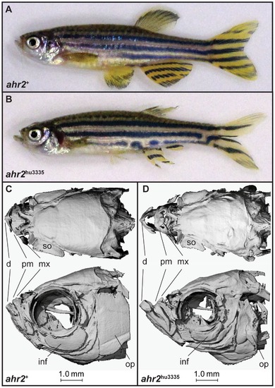
Fin and skeletal abnormalities observed in adult ahr2hu3335 zebrafish. A–B) Brightfield and (C–D) microCt imaging of adult ahr2+ and ahr2hu3335zebrafish. Notable differences were observed in the dentate (d), premaxilla (pm), maxilla (mx), supraorbital (so), infraorbital 3(inf) and operculum (op). PHENOTYPE:
|
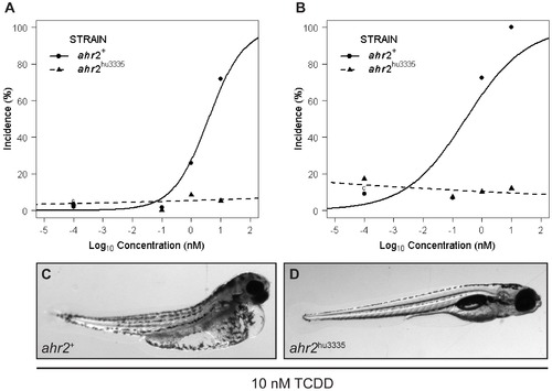
ahr2hu3335 embryos are resistant to TCDD-induced developmental abnormalities. A) Percent of embryos with axis malformations and B) percent incidence pericardial edema at 120 hpf in embryos treated with 0, 0.1, 1 or 10 nM TCDD from 6–24 hpf. Vehicle control groups (c, 0.1% DMSO) are displayed at 10-4 for graphing purposes. Data represent three independent experiments with 20 embryos per treatment group. C) Representative image of 120 hpf ahr2+ and (D) ahr2hu3335 embryos developmentally exposed to 10 nM TCDD. |

CYP1A protein expression patterns are ligand- and AHR isoform-dependent. CYP1A expression at 120 hpf in (A) ahr2+ and (B) ahr2hu3335 larvae following exposure to 1 nM TCDD from 6–24 hpf. C) Leflunomide-induced CYP1A expression at 72 hpf in wild-type and (D) ahr2hu3335 mutants. E) Leflunomide-induced CYP1A expression in AHR1B-morphant ahr2hu3335 larvae and F) ahr2hu3335 larvae co-injected with AHR1A and AHR1B morpholinos. (G) DMSO control. TCDD-exposed embryos were IHC processed side-by-side and imaged at 120 hpf using the same exposure settings and a single focal plane. Leflunomide-exposed embryos and DMSO control were processed side-by-side and imaged at 72 hpf using the same exposure times; images were created from a z-stack of 10 15.4 uM slices centered on the liver. |