
- Title
-
Inhibition of protein kinase A phenocopies ectopic expression of hedgehog in the CNS of wild-type and cyclops mutant embryos
- Authors
- Ungar, A.R., and Moon, R.T.
- Source
- Full text @ Dev. Biol.
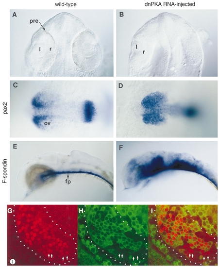
Inhibition of PKA phenocopies misexpression of hh. (A) Dorsal view (optical section) of a 28-hpf wild-type embryo. The lens (l) and pigmented retinal epithelium (pre) are evident. (B) Dorsal view (optical section) of a 28-hpf wild-type embryo injected with dnPKA RNA. Pigmented retinal epithelia have not developed; one lens is slightly reduced in size while the other has not developed. The lens was missing or reduced in size in 74% (n - 126) of eyes developing in dnPKA-injected embryos. Also, the ventricles of the brain fail to form properly in both dnPKA- and hh RNA-injected embryos. (C) pax2 RNA in a 14-hpf wild-type embryo (dorsal view). pax2 labels the anterior/proximal part of the optic vesicle (ov). (D) pax2 RNA in a 14-hpf embryo injected with dnPKA RNA (dorsal view). pax2 is expressed ectopically in the distal/posterior portion of the eye vesicle. In embryos injected with dnPKA RNA at the 1- to 8-cell stage, 89% (n - 46) displayed ectopic pax2 in the eye. (E) F-spondin RNA in a 28-hpf wild-type embryo (lateral view). Labeling is in the floor plate (fp). (F) F-spondin RNA in a 28-hpf embryo injected with dnPKA RNA (lateral view). Ectopic F-spondin was observed in 81% (n - 69) of dnPKA-injected embryos. (G) pax2 protein in the optic vesicle of a 15-hpf embryo which was injected with RNA encoding a dnPKA–GFP fusion protein. Anterior is to the upper left. The white dots roughly outline the extent of the optic vesicle. Labeling in the anterior/proximal half of the optic vesicle represents wild-type expression (compare to C) whereas labeling in the posterior/distal regions is ectopic. The white arrows indicate cells in which dnPKA–GFP was not detected (see H) but which express pax2 in ectopic regions of the eye vesicle. (H) dnPKA–GFP fusion protein in the same confocal section shown in G. dnPKA–GFP is primarily cytoplasmic. The white arrows indicate cells in which dnPKA–GFP was not detected but which express pax2 in ectopic regions of the eye vesicle (G). dnPKA–GFP was also not detected in these cells in adjacent z-series images taken 3 mm apart. (I) Superposition of G and H showing pax2 protein (red) and dnPKA-GFP (green). Key: fp, floor plate; l, lens; ov, optic vesicle; pre, pigmented retina epithelium; r, retina. EXPRESSION / LABELING:
|
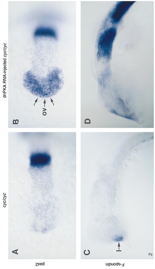
cyc mutant embryos are responsive to, but not rescued by, inhibition of PKA. cyc mutants injected with RNA encoding dnPKA–GFP fusion protein have dnPKA–GFP-containing cells in a fairly even but mosaic distribution (data not shown), as also seen in a wildtype background. (A) pax2 RNA in a 14-hpf cyc mutant embryo (dorsal view). (B) pax2 RNA in a 14-hpf cyc mutant embryo which was injected with dnPKA RNA (dorsal view). Although pax2 is expressed throughout the optic vesicle, the anterior– ventral fused morphology of the optic vesicle characteristic of cyc mutants is evident (arrows, and compare to Figs. 1C and 1D). (C) F-spondin RNA in a 30-hpf cyc mutant embryo (lateral view). F-spondin is absent from the ventral CNS (compare to Fig. 1E). The anterior F-spondin expression is in the lens and is also seen in wild-type embryos at this stage (data not shown). (D) F-spondin RNA in a 30-hpf cyc mutant embryo which was injected with dnPKA RNA (lateral view). Ectopic F-spondin is present in a pattern similar to that induced by dnPKA in wild-type embryos (compare to Fig. 1F). The cells in which F-spondin has been induced do not exhibit the characteristic morphology of the floor plate. Key: l, lens; ov, optic vesicle. EXPRESSION / LABELING:
|
Reprinted from Developmental Biology, 178(1), Ungar, A.R., and Moon, R.T., Inhibition of protein kinase A phenocopies ectopic expression of hedgehog in the CNS of wild-type and cyclops mutant embryos, 186-191, Copyright (1996) with permission from Elsevier. Full text @ Dev. Biol.