
- Title
-
Disruption of Eaat2b, a glutamate transporter, results in abnormal motor behaviors in developing zebrafish
- Authors
- McKeown, K.A., Moreno, R., Hall, V.L., Ribera, A.B., and Downes, G.B.
- Source
- Full text @ Dev. Biol.

tnt larvae exhibit an abnormal escape response at 48 hpf. Selected frames from high-speed video recordings are shown with times indicated in each frame in milliseconds. (A) A wild-type larva exhibits a normal C-bend (A2 — asterisk) in response to a touch stimulus, followed by smaller-amplitude undulations (A3–A12). (B) A tnt mutant abnormally exhibits C-bends (asterisks) throughout the duration of the escape response (B3, B8, and B10). PHENOTYPE:
|
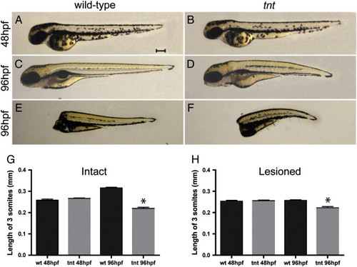
tnt larvae demonstrate paralysis and body shortening at 96 hpf, which can occur in the absence of brain input. Lateral views are shown of wild-type and tnt larvae and lesioned preparations. (A,B) Wild-type and tnt larvae are of similar length along the rostral–caudal axis at 48 hpf. Scale bar = 0.2 mm, applies to panels A–F. (C,D) At 96 hpf, tnt mutants are shorter than wild-type siblings. (E,F) At 96 hpf, tnt lesioned preparations, in which brain input was removed at 46 hpf, are shorter than wild-type, suggesting that the brain is not required for this aspect of the tnt phenotype. Quantification of a three-somite length is shown for (G) intact and (H) lesioned larvae (n = 8, p < 0.05). PHENOTYPE:
|

slc1a2b knockdown phenocopies tnt mutants at 48 and 96 hpf. Kinematic traces are shown, with 0° indicating a straight body and positive and negative angles representing body bends in opposite directions. Time is shown in seconds. Five representative traces are shown for each condition. (A) Wild-type larvae exhibit one large amplitude bend, a C-start, followed by smaller amplitude body bends. (B) tnt larvae exhibit several large amplitude body bends throughout the escape response. (C) Embryos injected with the standard control morpholino are similar to wild-type. (D) slc1a2b morphant larvae exhibit many large amplitude C-bends similar to tnt mutants. (E) Quantification of the number of large amplitude body bends (defined as greater than 110°) observed within an escape. Both tnt mutant and slc1a2b morphant larvae perform significantly more large amplitude body bends (n = 20, p < 0.05). Asterisks indicate significant differences. (F) tnt mutant and slc1a2b morphant larvae also performed significantly longer escape responses (n = 20, p < 0.05). (G) Embryos injected with the tnt morpholino also demonstrate body shortening along the rostral-caudal axis at 96 hpf, but not 48 hpf, compared to embryos injected with the control morpholino (n = 12 to 14, p < 0.01). |
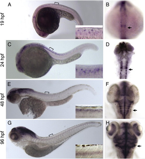
slc1a2b exhibits dynamic expression in the hindbrain and spinal cord across development. Lateral (A,C,E,G) and dorsal views (B,D,F,H) are shown, with the brackets indicating the regions shown at higher magnification in the insets. For orientation in dorsal images, the arrows indicate the location of the otic placode. (A,B) At 19 hpf, slc1a2b shows robust expression in rhombomere 4 of the hindbrain with punctate, less pronounced expression in the spinal cord. (C,D) At 24 hpf, slc1a2b expression continues to expand throughout the hindbrain and caudally into the spinal cord compared to 19 hpf. (E,F) At 48 hpf, slc1a2b is expressed in the hindbrain and in the spinal cord. (G,H) At 96 hpf, slc1a2b is widely expressed in the hindbrain and spinal cord, other brain regions, and in the retina. EXPRESSION / LABELING:
|
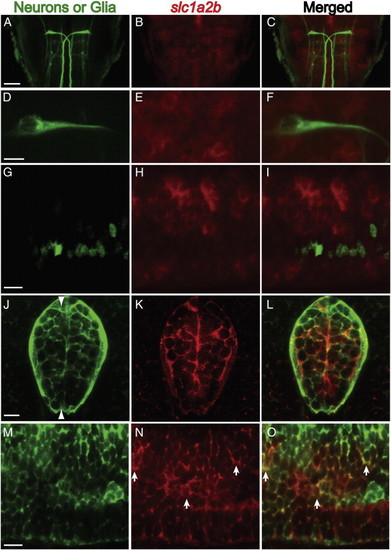
slc1a2b mRNA is expressed in glial cells at 48 hpf. (A) The 3A10 antibody labels the Mauthner cell in the hindbrain. The scale bar = 50 μm. (B) slc1a2b mRNA is expressed in the hindbrain in cells around the Mauthner cell. (C) The merged images show that slc1a2b mRNA is not detected within the Mauthner cell. (D–F) Higher magnification views of a Mauthner cell, slc1a2b staining, and the merged images. The scale bar in this and all following panels = 10 μm. (G–I) Lateral views of Tg(mnx1:GFP) larvae, which express GFP in motor neurons and some ventral interneurons, stained for slc1a2b mRNA expression. Little colocalization between GFP+ neurons and slc1a2b mRNA was observed. (J–K) Cross-sectional views of the spinal cord of Tg(gfap:GFP) transgenic larvae. GFAP is expressed predominantly in glial cells at this stage of development. slc1a2b expression was detected in some GFAP-positive cells. (M–O) Lateral views of Tg(gfap:GFP) larvae stained for slc1a2b mRNA expression. The arrowheads in J indicate the plane in which the lateral views are taken. The arrows indicate examples of a GFP+ cell in which slc1a2b expression was detected. Similar results were obtained using 96 hpf larvae. EXPRESSION / LABELING:
|
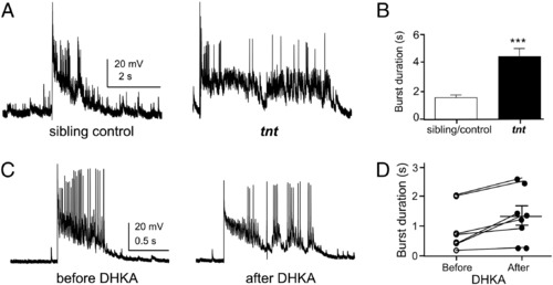
Motor neurons in tnt mutants exhibit increased burst duration. (A,B) Supraspinal electrical stimulation was used to elicit bursts in CaP motor neurons to evaluate synaptic input in 72–96 hpf larvae. Burst duration was increased in tnt CaP neurons compared to that of sibling controls (for sibling control and tnt embryos, 8 and 6 cells were analyzed, respectively; p = 0.0001, two-tailed t-test). (C,D) To further assess the effects of decreased Eaat2 function, stimulated bursts were recorded before and after addition of the selective blocker DHKA. Addition of DHKA (160 µM) significantly increased burst duration (p = 0.004, two-tailed pair t-test), similar to the effect of the tnt mutation. PHENOTYPE:
|

Unillustrated author statements |
Reprinted from Developmental Biology, 362(2), McKeown, K.A., Moreno, R., Hall, V.L., Ribera, A.B., and Downes, G.B., Disruption of Eaat2b, a glutamate transporter, results in abnormal motor behaviors in developing zebrafish, 162-171, Copyright (2012) with permission from Elsevier. Full text @ Dev. Biol.