
- Title
-
Roles of Hedgehog pathway components and retinoic acid signalling in specifying zebrafish ventral spinal cord neurons
- Authors
- England, S., Batista, M.F., Mich, J.K., Chen, J.K., and Lewis, K.E.
- Source
- Full text @ Development
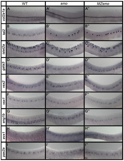
V0v, V1 and V2 cells persist in the absence of Hh signalling. (A-I3) In situ hybridisation for the indicated marker genes in lateral views of 24-hpf trunk of WT, smo mutant and MZsmo mutant zebrafish embryos. Asterisks indicate tal2-expressing cells (B′,B′′) and islet2a-expressing MNs (C′,C′′) that still form in smo and MZsmo mutants. islet2a is also expressed in more dorsal Rohon-Beard cells (arrowheads in C-C′′). Rohon-Beard cells are unaffected in smo or MZsmo mutants (C′,C′′). eng1b is also expressed in muscle pioneer cells ventral to the spinal cord in WT embryos (arrowheads in G). These cells are absent in smo or MZsmo mutants (G′,G′′). Scale bar: 50 μm. |
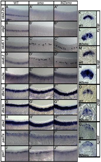
Ventral progenitor domains are lost at 24 hpf in the absence of Hh signalling. (A-J′′) Lateral views of zebrafish trunk and (K-R) transverse sections at 24 hpf. Asterisks (C′,C′′,D′,D′′) indicate weakly expressing cells. Scale bar: 50 μm in A-J′′; 40 μm in K-R. |
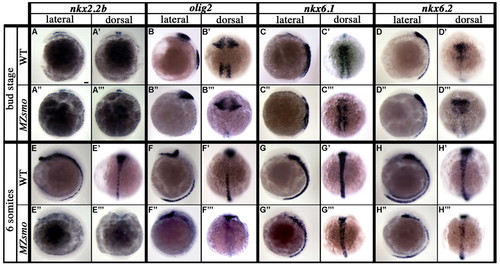
nkx6.1, nkx6.2 and olig2 are expressed in the spinal cord at early somitogenesis stages in the absence of Hh signalling. (A-H′′ ′) Lateral and dorsal views of nkx2.2b, olig2, nkx6.1 and nkx6.2 expression in WT and MZsmo mutant zebrafish embryos at bud stage (10 hpf) and 6 somites (12 hpf). Scale bar: 50 μm. |
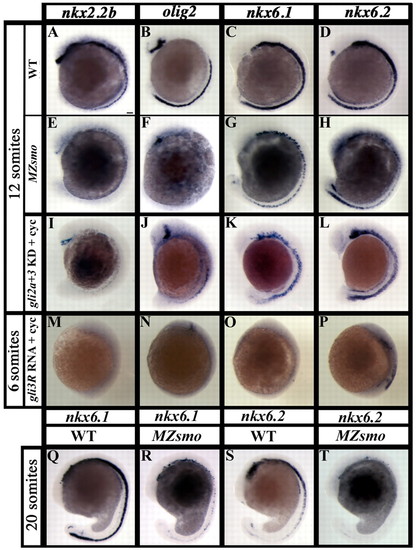
Progenitor domain expression at somitogenesis stages. (A-T) Lateral views of WT zebrafish embryos (A-D,Q,S), MZsmo mutants (E-H,R,T) and embryos treated with cyclopamine (cyc) and injected with gli2a, gli3 and p53 MOs (gli2a + 3 KD; I-L) or gli3R RNA (M-P). When gli2a and gli3 were knocked down, nkx2.2b was absent (n=23; I), a small amount of olig2 expression was recovered (n=25; J), and nkx6.1 (n=22; K) and nkx6.2 (n=19; L) expression was much stronger in all injected embryos. When gli3R RNA was injected, all embryos completely lacked nkx2.2b (n=21; M) and olig2 (n=45; N) expression. nxk6.1 expression was lost in 54% of embryos; the remaining embryos had strongly reduced staining (n=28; O). nkx6.2 expression was lost in 8%, strongly reduced in 81%, and less reduced in 11% of injected embryos (n=26; P). Scale bar: 50 μm. |
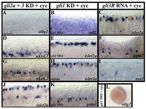
Manipulating Gli activity in embryos with abrogated Hh signalling. (A-L) Lateral views of spinal cord expression in 24-hpf zebrafish embryos treated with cyclopamine (cyc) and injected with either gli2a, gli3 and p53 MOs (A,D,G,J), gli1 and p53 MOs (B,E,K) or gli3R RNA (C,F,I), a 24-hpf gli1 mutant (detour) embryo treated with cyclopamine (H) and a 6-somite (12-hpf) embryo treated with cyclopamine and injected with gli1 and p53 MOs (L). For L, 28/29 injected embryos had a complete lack of spinal cord olig2 expression; the remaining embryo had a very small amount of spinal cord staining. Scale bar: 50 μm in A-K; 250 μm in L. |
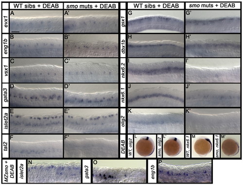
Postmitotic and progenitor domain markers are reduced in the absence of RA signalling. Lateral views of WT sibling, smo mutant and MZsmo mutant 24-hpf spinal cords (A-K′,N-P) and WT sibling and smo mutant 6-somite (12-hpf) embryos (L-M′) treated with DEAB. Scale bar: 50 μm in A-K′,N-P; 250 μm in L-M′. |
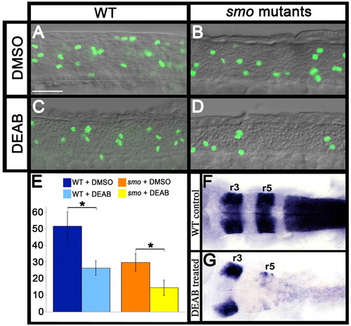
DEAB treatment causes a reduction in cell proliferation. (A-D) Lateral views of phospho-Histone H3 staining in DMSO-treated control and DEAB-treated spinal cords of WT zebrafish embryos and smo mutants at 24 hpf. Scale bar: 50 μm. (E) Average number (n=5 embryos) of phospho-Histone H3-positive cells in spinal cord adjacent to somites 6-10. Statistically significant differences between embryos of the same genotype are indicated (*, P<0.05, Student’s t-test). Error bars denote s.d. (F,G) dorsal views of hindbrain showing dramatically reduced egr2b expression in rhombomere (r) 5 and the loss of hoxb4a expression in the anterior spinal cord of DEAB-treated embryos (G). |
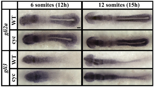
gli2a and gli3 expression. Dorsal flat-mount views of gli2a and gli3 expression in WT and cyclopamine-treated (cyc) embryos. Neither gene is obviously expressed in ventral spinal cord at 6 somites but there might be expression in the ventral spinal cord by 12 somites. For gli3, at least, this expression appears to be stronger in embryos treated with cyclopamine. Scale bar: 50 μm |