
- Title
-
Genetic Association for Renal Traits among Participants of African Ancestry Reveals New Loci for Renal Function
- Authors
- Liu, C.T., Garnaas, M.K., Tin, A., Kottgen, A., Franceschini, N., Peralta, C.A., de Boer, I.H., Lu, X., Atkinson, E., Ding, J., Nalls, M., Shriner, D., Coresh, J., Kutlar, A., Bibbins-Domingo, K., Siscovick, D., Akylbekova, E., Wyatt, S., Astor, B., Mychaleckjy, J., Li, M., Reilly, M.P., Townsend, R.R., Adeyemo, A., Zonderman, A.B., de Andrade, M., Turner, S.T., Mosley, T.H., Harris, T.B., Rotimi, C.N., Liu, Y., Kardia, S.L., Evans, M.K., Shlipak, M.G., Kramer, H., Flessner, M.F., Dreisbach, A.W., Goessling, W., Cupples, L.A., Kao, W.L., Fox, C.S.
- Source
- Full text @ PLoS Genet.
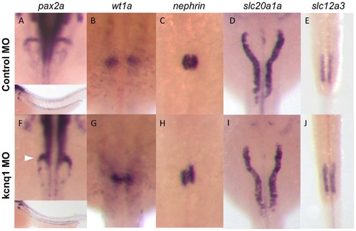
Kcnq1 knockdown in zebrafish embryos causes glomerular abnormalities. (A–E) Zebrafish embryos injected with control morpholino show normal glomerular and tubular morphology, as shown by in situ hybridization for the global kidney marker pax2a (A, inset showing lower-magnification image, with staining in both glomerulus and tubules), the podocyte markers wt1a (B) and nephrin (C), and the proximal and distal tubular markers slc20a1a (D) and slc12a3 (E). (F–J) Injection of the kcnq1 antisense morpholino, targeting the ATG site of the gene, at the one-cell stage results in significant changes in glomerular gene expression (F–H). No changes were observed in the proximal tubule (I) or the distal tubule (J). |
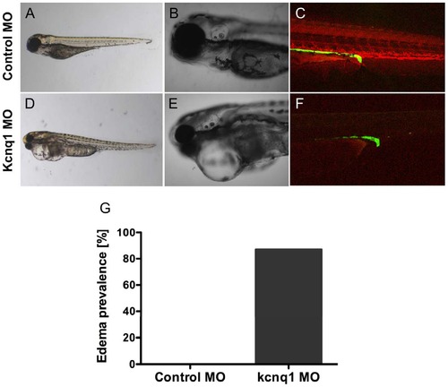
Tg(cdh17:GFP) embryos were injected with 200 uM control or kcnq1 morpholino (MO) at the 1-cell stage. Embryos were subsequently injected in the sinus venosus with 10,000 MW rhodamine dextran at 48 hpf. Dextran clearance was monitored over the next 48 hours by confocal microscopy. Compared to control MO-injected embryos (A–C), kcnq1 MO-injected embryos exhibit edema (D, E) and increased dextran clearance (F) 48 hours post-injection (hpi), suggestive of a filtration defect. At 5 dpf, 87% of kcnq1 morphant embryos (n = 38) develop edema (G). PHENOTYPE:
|

Analysis of glomerular architecture after kcnq1 knockdown by electron microscopy does not reveal significant changes. (a–c) Glomerular architecture at 120 hpf after injection of control morpholino visualized by electron microscopy at 8000-, 15,000, and 50,000-fold magnification reveals endothelial capillaries (C) with basement membrane (BM), podocytes (P) and foot processes (FP) similar to mammalian glomerular ultrastructure. (d–f) Transient knockdown of kcnq1 does not significantly change glomerular anatomy. PHENOTYPE:
|
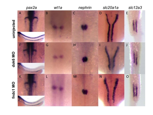
Dok6 and fndc1 knockdown in zebrafish embryos does not affect kidney development. (a–e) Uninjected control zebrafish embryos show normal glomerular and tubular morphology, as shown by in situ hybridization for the global kidney marker pax2a (a, inset showing lower-magnification image, with staining in both glomerulus and tubules), the podocyte markers wt1a (b) and nephrin (c), and the proximal and distal tubular markers slc20a1a (d) and slc12a3 (e). (f–o) Injection of dok6 (f–j) or fndc1 (k–o) morpholinos at the one-cell stage results in no significant changes in glomerular or tubular gene expression. EXPRESSION / LABELING:
|
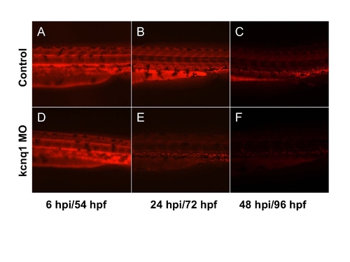
kcnq1 knockdown enhances loss of fluorescent dextran. Control and kcnq1 MO injected embryos were loaded with rhodamine-labeled dextran at 48 hpf. >80 embryos were analyzed for each group in 3 separate experiments (a,d) Fluorescence microscopy at 6 hpi reveals equal fluorescence loading between embryos. (b,e) At 72 hpf (24 hpi), fluorescence intensity in kcnq1 morphants is diminished. (c,f) At 96 hpf (48 hpi) control embryos retain their fluorescence, but kcnq1 morphants have significantly diminished fluorescence. |