
- Title
-
Eriocaulon buergerianum extract protects PC12 cells and neurons in zebrafish against 6-hydroxydopamine-induced damage
- Authors
- Wang, M., Zhang, Z., Cheang, L.C., Lin, Z., and Lee, S.M.
- Source
- Full text @ Chin. Med.
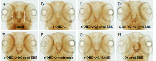
EBE recovered 6-OHDA-induced dopaminergic neuron loss in zebrafish. (A-H) 2 dpf zebrafish was exposed to 250 μM 6-OHDA and different concentrations of EBE or 100 μM Nomifensin or 100 μM L-NAME or 100 μg/mL EBE for 24 hours, except the control. The viability of dopaminergic neurons of the zebrafish (indicated by white brackets) was evaluated with anti-tyrosine hydroxylase (TH) immunostaining. Ventral view: anterior to the top. |
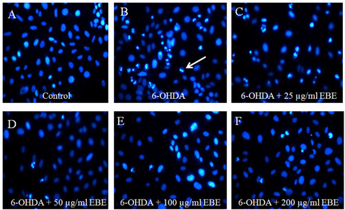
EBE reduced apoptosis induced by 6-OHDA in PC12 cells. Cells were stained with DNA-binding fluorescent dye Hoechst 33342. (A) Control: untreated group; (B) 6-OHDA-treated group (1 mM, 8 hours): chromatin condensation and DNA fragmentation were indicated by the white arrows; (C-F) EBE-pretreated groups (25, 50, 100 and 200 μg/mL respectively, 12 hours), followed by 6-OHDA exposure (1 mM, 8 hours): less apoptotic bodies were identified, colony reduction and cell shrinkage induced by 6-OHDA were also reversed. |
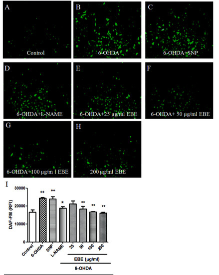
EBE inhibited 6-OHDA-induced nitric oxide (NO) over-production in PC12 cells. PC12 cells were pretreated with or without 250 μM L-NAME, 100 μM SNP and 25, 50, 100 and 200 μg/mL EBE for 12 hours, then exposed to1 mM 6-OHDA for another hour. (A-H) Intracellular NO was identified using fluorescent indicator, DAF-FM diacetate; (I) The NO fluorescent intensity was quantified by a multi-label counter. ++ P = 0.008 versus control group (without 6-OHDA treatment); * P = 0.044 versus OHDA group; ** P = 0.005 versus OHDA group. All experiments were repeated 3 times. |