
- Title
-
Early embryonic gene expression profiling of zebrafish prion protein (prp2) morphants
- Authors
- Nourizadeh-Lillabadi, R., Seilø Torgersen, J., Vestrheim, O., König, M., Aleström, P., and Syed, M.
- Source
- Full text @ PLoS One
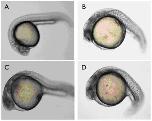
Larvae were immobilized in CyGEL prior to microscopy. A) Non-injected wt control. B–D) three individual morphants displaying defective midbrain and hindbrain development. PHENOTYPE:
|
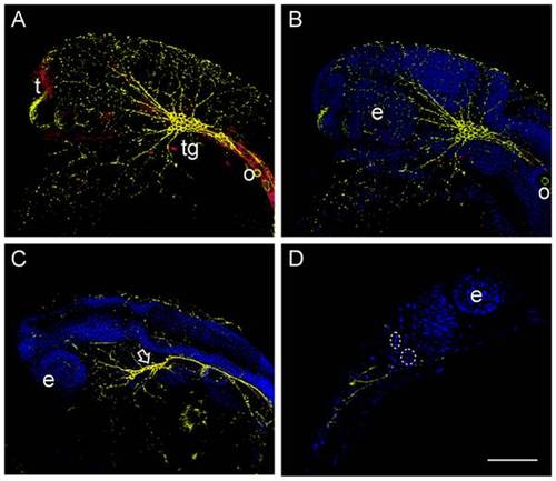
Neuronal structures were visualized by the Zn12 antibody (Yellow), whereas Prp2 was detected with a rabbit anti serum (Red). A) In the wild type 24 hpf Prp2 is observed in the trigeminal ganglion and telencephalon, but also present in other neuronal tissue. B) Neuronal structures visualized by HNK-1 staining (Zn-12 antibody) in the same specimen as in A. C) Aberrant morphology of the trigeminal ganglion in Prp2 morphants (arrow) as well as reduced number of peripheral neurons visualized by HNK-1 staining. D) Non deconvolved single plane image of a morphant with condensed nuclei (inside stippled ring). Abbreviations: eye (e), telencephalon (t) and trigeminal ganglion (tg). |