
- Title
-
Zebrafish teeth as a model for repetitive epithelial morphogenesis: dynamics of E-cadherin expression
- Authors
- Verstraeten, B., Sanders, E., van Hengel, J., and Huysseune, A.
- Source
- Full text @ BMC Dev. Biol.

Expression pattern of E-cadherin in first-generation teeth. Left panels: mRNA expression; middle panels: protein expression; right panels: diagram of different cell layers of the tooth. A, B, C: Initiation phase. D, E, F: Morphogenesis phase; G, H, I: Early cytodifferentiation and attachment phase; J, K, L: Late cytodifferentiation phase and initiation of successor. ph.c.: pharyngeal cavity; ph.e.: pharyneal epithelium; *: dental papilla; arrowhead: enamel organ; block arrow: initiation replacement tooth. Diagrams: green cells: pharyngeal epithelium; red cells: outer dental epithelium; blue cells: inner dental epithelium. Scale bars = 2 5 μm. |
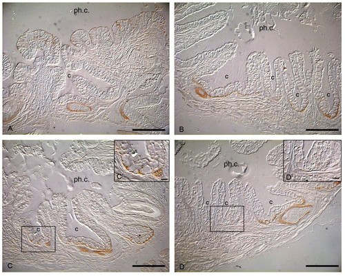
Uneven distribution of E-cadherin in the crypt epithelium. Sections of whole mount immuocytochemical examination of dissected adult jaw. The rostral crypts in the pharyngeal region of adult zebrafish express E-cadherin throughout the epithelium (A). This contrasts to the more caudal crypt epithelium, which loses E-cadherin expression, except along the crypt bases (B). Cells placed rostrally at the base of the crypt always have stronger E-cadherin expression (C, C′) than cells placed more caudally within the same crypt (D, D′). c: crypt; ph.c.: pharyngeal cavity. Scale bars = 50 um, scale bar C′, D′ = 10 μm. |

E-cadherin expression pattern during tooth replacement. Sections of whole mount immunocytochemical examination of dissected adult jaw. A, A′: Successional dental lamina is positive for E-cadherin; B: Morphogenesis phase; C: Early cytodifferentiation, IDE and ODE express E-cadherin; D: Late cytodifferentiation. *: dental papilla; arrowhead: enamel organ; c: crypt slightly posterior to the tip of the functional predecessor; L: lateral; M: medial. Scale bars = 50 μm, scale bar A′ = 25 μm. |

Beta-catenin immunolocalization in the successional lamina. The onset of formation of a replacement tooth, the successional lamina, has strong p-catenin expression along the membrane of every cell (A, A′). This β-catenin expression co-localizes with the expression of E-cadherin suggesting that the cadherin-catenin complex is functional as adhesive force. arrowhead: successional lamina; c: crypt surrounding the tip of the functional predecessor. Scale bar = 50 μm, scale bar A′ = 15 μm. |