
- Title
-
Caffeine-induced effects on heart rate in zebrafish embryos and possible mechanisms of action: an effective system for experiments in chemical biology
- Authors
- Rana, N., Moond, M., Marthi, A., Bapatla, S., Sarvepalli, T., Chatti, K., and Challa, A.K.
- Source
- Full text @ Zebrafish
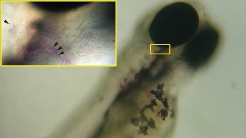
Digital still images of live 2-dpf zebrafish embryos at 10x and 40x (inset) objective magnifications, stained with aqueous solutions of ruthenium red. Arrowheads point to individual cells with ruthenium red staining. |
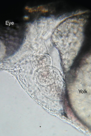
Digital still image of a 2-day postfertilization (dpf ) zebrafish embryo heart at 40x objective magnification, showing the two chambers of the heart with the valve ring in the middle. The larger chamber is the ventricle and the smaller chamber is the atrium. The yolk sac is visible adjacent to the heart. Red blood corpuscles are discernible within the heart. |
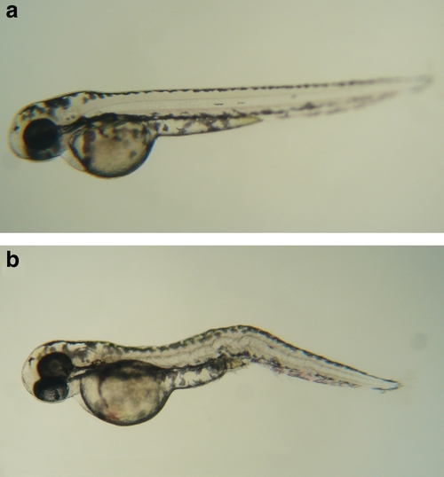
Digital still images of 2-dpf zebrafish embryos at 4x objective magnification. (a) The appearance of a normal anesthetized embryo without caffeine treatment; (b) an embryo treated with 10mM caffeine, showing the characteristic kinking in the trunk/tail region. |